A method for adaptively extracting an MR image of a cervical tumor

An adaptive, image-based technology, applied in the field of medical image processing, can solve the problems that doctors cannot accurately determine the boundaries of cervical tumors, do not automatically extract cervical tumor areas, and medical image noise, so as to reduce human subjective bias and omit manual parameter adjustment The cumbersome, high clinically significant effect of

Inactive Publication Date: 2019-04-16
NORTHEASTERN UNIV
View PDF2 Cites 3 Cited by
  • Summary
  • Abstract
  • Description
  • Claims
  • Application Information

AI Technical Summary

Problems solved by technology

However, due to the influence of imaging equipment, patient body movement and other factors, there will be noise and artifacts in medical images, making it difficult for doctors to accurately determine the boundaries of cervical tumors.
Currently, there are no studies on the automatic extraction of cervical tumor regions from MR images

Method used


Image

  • A method for adaptively extracting an MR image of a cervical tumor
  • A method for adaptively extracting an MR image of a cervical tumor
  • A method for adaptively extracting an MR image of a cervical tumor

Examples


Embodiment Construction

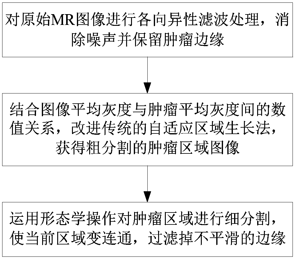
[0037] Such as figure 1 As shown, a kind of adaptive extraction method of cervical tumor MR image of the present invention comprises the following steps:

[0038] Step 1: Anisotropic filtering is performed on the original MR image to eliminate noise and retain tumor margins. The step 1 includes:

[0039] Step 1.1: Convert the Dicom format file provided by the hospital into the original MR image in raw format, which is convenient for computer reading and subsequent further processing;

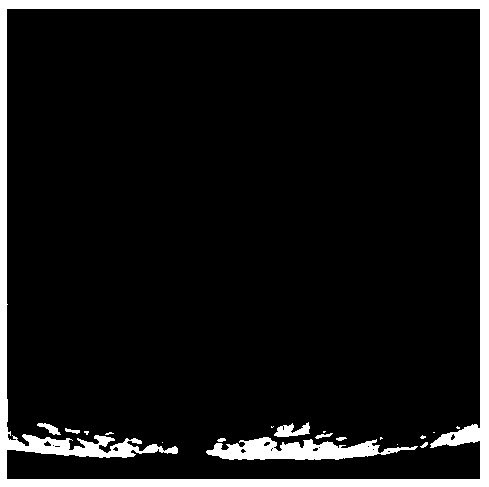
[0040] Step 1.2: intercept the rectangular part containing the cervical region from the original MR image, and use the rectangular region as the basis for segmentation, such as Figure 2a shown;

[0041] Step 1.3: Anisotropic filtering is performed on the segmented image to effectively remove noise and preserve tumor edges.

[0042] Anisotropic filtering is a method that can both remove noise and preserve image edges. For images, anisotropy means that the gradient changes in the four directi...


PUM


Abstract

The invention relates to the technical field of medical image processing, and provides a method for adaptively extracting an MR image of a cervical tumor, and the method comprises the steps: 1, carrying out the anisotropic filtering of an original MR image, eliminating noise, and keeping the edge of a tumor; Step 2, for the filtered MR image, setting a proportionality coefficient in combination with the numerical relationship between the average gray scale of each image and the average gray scale of the tumor, and improving a traditional adaptive region growth method to obtain a roughly segmented tumor region image; And step 3, performing fine segmentation on the tumor region by using morphological operation to enable the current region to be communicated and smooth the edge. According tothe method, an improved adaptive region growth method is utilized, the coincidence rate of an obtained segmentation result and a tumor region gold standard manually drawn by a doctor is high, the complexity of manually setting a threshold value is avoided, the artificial participation degree is reduced, artificial subjective deviation is reduced, universality is achieved, and a reasonable reference is provided for clinical diagnosis.

Description

technical field [0001] The invention belongs to the field of medical image processing, and relates to a method for adaptively extracting MR images of cervical tumors. Background technique [0002] Cervical cancer is one of the common gynecological malignancies, accounting for more than half of female genital cancers. In recent years, the morbidity and mortality of cervical cancer have increased significantly, and the prevention of cervical diseases and the screening of cervical cancer have also been paid more and more attention by the majority of women. With the development of medical imaging technology, MRI technology is more and more widely used in the diagnosis and treatment of cervical cancer. Doctors draw the boundaries of cervical tumors on MR images based on their own experience, and then judge the stage of cervical cancer. However, due to the influence of imaging equipment, patient body movement and other factors, medical images will have noise and artifacts, makin...

Claims


Application Information

Patent Timeline
Patent Type & Authority Applications(China)
IPC IPC(8): G06T7/187G06T5/00
CPCG06T5/002G06T7/187G06T2207/30096G06T2207/10088
Inventor 苏缨韩芳芳孙伟中马贺康颖
Owner NORTHEASTERN UNIV
Why Eureka
Try Eureka