Supercharge Your Innovation With Domain-Expert AI Agents!

Apparatus for tubulus detection from a tissue biopsy

A technology for tissue biopsy and tubular objects, applied in image analysis, image enhancement, instruments, etc., can solve problems such as complex detection systems, and achieve the effect of less material loss and more material loss

Inactive Publication Date: 2019-04-16
KONINKLJIJKE PHILIPS NV
View PDF16 Cites 0 Cited by
  • Summary
  • Abstract
  • Description
  • Claims
  • Application Information

AI Technical Summary

Problems solved by technology

Requires complex, exotic and expensive detection systems, e.g., those based on confocal microscopy and OPT techniques

Method used


Image

  • Apparatus for tubulus detection from a tissue biopsy
  • Apparatus for tubulus detection from a tissue biopsy
  • Apparatus for tubulus detection from a tissue biopsy

Examples


Embodiment Construction

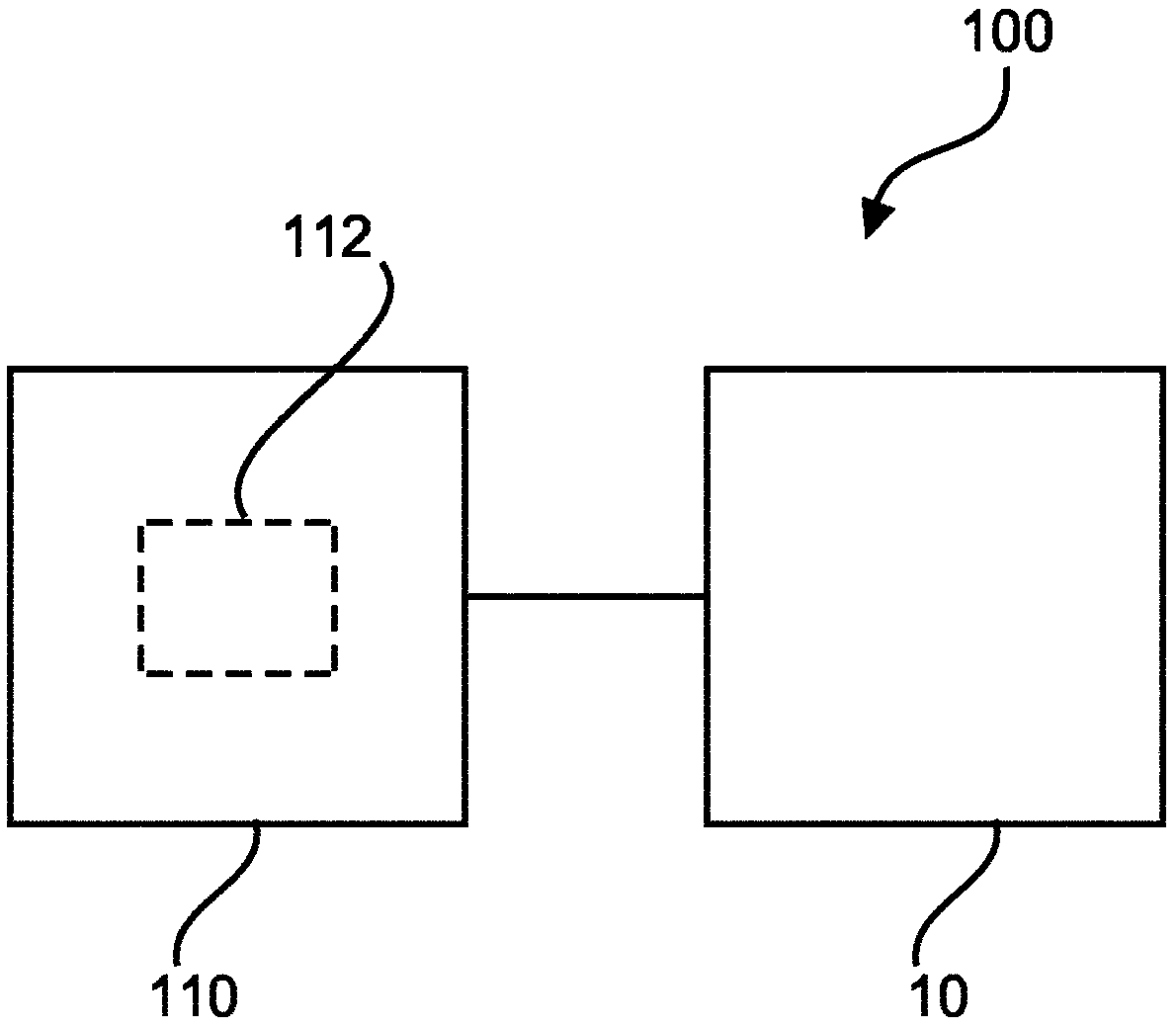
[0070] figure 1 A device 10 for detecting tubing from a tissue biopsy is shown. The device 10 includes an input unit 20 , a processing unit 30 and an output unit 40 . The input unit 20 is configured to provide the processing unit 30 with a plurality of 2D images of the complete tissue biopsy. Each 2D image corresponds to a different depth location in the complete tissue biopsy, and each 2D image includes image data of the complete tissue biopsy. The processing unit 30 is configured to determine a measure of the local intensity variation of the image data of the intact tissue biopsy in the region of the at least one 2D image. The processing unit 30 is further configured to locate at least part of the tubular in the region of the at least one 2D image based on the determined measure of the local intensity variation. The localization comprises determining, in the region of the at least one 2D image, where the measure of the local variation of the intensity is below a threshold...


PUM


Abstract

The present invention relates to an apparatus (10) for tubulus detection from a tissue biopsy. It is described to provide (210) a plurality of 2D images of a tissue biopsy, wherein each 2D image corresponds to a different depth position in the tissue biopsy, and wherein each 2D image comprises image data of the tissue biopsy. A measure of a local variation of intensity is determined (220) in the image data of the tissue biopsy in a region of at least one 2D image. At least part of a tubulus is located (230) in the region of the at least one 2D image on the basis of the determined measure of the local variation of intensity. The locating (230) involves determining (240) locations in the region of the at least one 2D image where the measure of the local variation in intensity is below a threshold. Data representative of the location of the at least part of the tubulus in the region of the at least one 2D image are output (250).

Description

technical field [0001] The invention relates to a device for detecting tubing from a tissue biopsy, a system for detecting tubing from a tissue biopsy, a method for detecting tubing from a tissue biopsy, and a computer program element and a computer readable medium. Background technique [0002] Analyzing tissue using 2D pathology requires that tissue samples be sliced ​​into thin sections approximately 4 μm thick and stained to increase image contrast. More recently, 3D pathology has been developed, enabling better visualization and analysis of, for example, growth patterns of cancer. However, detecting tubular features in images is based on using the color intensity of the stain applied to the tissue biopsy and using exotic microscopy techniques such as confocal microscopy and optical projection tomography. [0003] E.g: [0004] "Structural and molecular interrogation of intact biological systems" by K. Chung et al. (Nature 497, p. 332, May 2013) describe a method for ...

Claims


Application Information

Patent Timeline
Patent Type & Authority Applications(China)
IPC IPC(8): G06T7/12
CPCG06T2207/10056G06T2207/30024G06T2207/30081G06T7/12G06T7/0012
Inventor C·韦雷坎普P·J·范德扎格M·J·A·阿塞尔曼
Owner KONINKLJIJKE PHILIPS NV
Features
  • R&D
  • Intellectual Property
  • Life Sciences
  • Materials
  • Tech Scout
  • Unparalleled Data Quality
  • Higher Quality Content
  • 60% Fewer Hallucinations
Learn More