
A method and system for measuring core infarction volume based on head CT images

A CT imaging and skull technology, applied in the field of medical imaging and computer, can solve the problems of accuracy, repeatability and objectivity limitations, affecting measurement results, loss of information, etc., to eliminate or reduce the interference of subjective factors, accurate measurement Effect

Active Publication Date: 2021-02-19
UNION STRONG (BEIJING) TECH CO LTD
View PDF4 Cites 0 Cited by
  • Summary
  • Abstract
  • Description
  • Claims
  • Application Information

AI Technical Summary

Problems solved by technology

Among them, the key to manually measuring the core infarct volume is to manually outline the infarct focus, which is very cumbersome and has great limitations in accuracy, repeatability, and objectivity.
Computer-aided measurement of core infarct volume mostly uses the method of image segmentation for measurement, but this method will lose some information in the process of processing, which will affect the measurement results

Method used


Image

  • A method and system for measuring core infarction volume based on head CT images
  • A method and system for measuring core infarction volume based on head CT images
  • A method and system for measuring core infarction volume based on head CT images

Examples


Embodiment Construction

[0071] In order to enable those skilled in the art to better understand the technical solutions in this specification, the technical solutions in the embodiments of this specification will be clearly and completely described below in conjunction with the drawings in the embodiments of this specification. Obviously, the described The embodiments are only some of the embodiments of the present application, but not all of them. Based on the embodiments of this specification, all other embodiments obtained by persons of ordinary skill in the art without creative efforts shall fall within the scope of protection of this application.

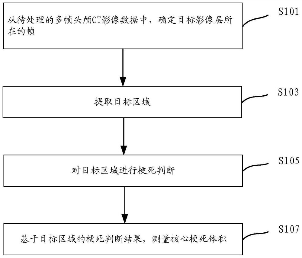
[0072] figure 1 A frame diagram of a method for measuring core infarct volume based on head CT images provided in the embodiment of this specification, the specific steps include:

[0073] Step S101: From the multiple frames of head CT image data to be processed, respectively determine the frame where the target image layer is located.

[0074] A CT...


PUM


Abstract

The embodiment of this specification discloses a method and system for measuring core infarct volume based on cranial CT images, belonging to the field of medical imaging. The embodiment of this specification solves the problems of large subjectivity differences and poor accuracy of core infarction volume measurement results in the prior art through the measurement method of core infarction volume based on head CT images. The measurement method includes: determining the frame where the target image layer is located from the multi-frame head CT image data to be processed; extracting the target area; performing infarction judgment on each target area included in the target area, and outputting the infarction judgment result; Regional infarct judgment results, measuring the core infarct volume. The measurement method and system provided in the embodiments of this specification can quickly and accurately measure the core infarct volume, eliminate or reduce the interference of subjective factors, and serve as a computer-aided method to provide objective evidence for stroke and other medical research.

Description

technical field [0001] This specification relates to the fields of medical imaging and computer technology, and in particular to a method and system for measuring core infarct volume based on head CT images. Background technique [0002] Core infarction is one of the important indicators in the evaluation of acute ischemic stroke (AIS). Among them, core infarction refers to the brain tissue with irreversible damage, which refers to the area where the cerebral blood flow decreases by more than 30% compared with normal brain tissue, and it is displayed as low density area. The size of the infarct core is closely related to the clinical prognosis of patients. The smaller the infarct core, the greater the likelihood that the patient will have a good prognosis. At the same time, evaluation of the infarct core can also predict the risk of complications of endovascular therapy. Therefore, accurate evaluation of the infarct core is helpful for screening stroke patients who are su...

Claims


Application Information

Patent Timeline
Patent Type & Authority Patents(China)
IPC IPC(8): G06T7/00G06T7/62
CPCG06T7/0012G06T2207/10081G06T2207/30016G06T7/62
Inventor 金海岚宋凌印胤杨光明秦岚
Owner UNION STRONG (BEIJING) TECH CO LTD
Learn More