An image screening method and device

An image and screening technology, applied in image analysis, image data processing, instruments, etc., can solve problems such as chest X-ray disease diagnosis difficulties, organ occlusion, etc., and achieve the effect of reducing bone occlusion and high accuracy

Active Publication Date: 2019-05-17
SHANGHAI UNITED IMAGING INTELLIGENT MEDICAL TECH CO LTD
View PDF7 Cites 5 Cited by
  • Summary
  • Abstract
  • Description
  • Claims
  • Application Information

AI Technical Summary

Problems solved by technology

However, chest radiograph is a 2D image generated by X-ray irradiation in a single direction, and there are se

Method used


Image

  • An image screening method and device
  • An image screening method and device
  • An image screening method and device

Examples


Embodiment 1

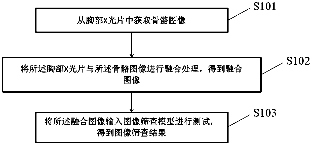
[0084] Such as figure 1 As shown, this embodiment provides a schematic flow chart of an image screening method, and this specification provides the method operation steps described in the embodiment or flow chart, but based on routine or non-creative work, it may include more or less operation steps. The sequence of steps enumerated in the embodiments is only one way of execution sequence of many steps, and does not represent the only execution sequence. Specifically as figure 1 As shown, the method includes:

[0085] S101. Acquiring bone images from chest X-ray films;

[0086] S102. Perform fusion processing on the chest X-ray film and the bone image to obtain a fusion image;

[0087] S103. Input the fused image into an image screening model for testing, and obtain an image screening result; the image screening model includes a model determined by deep learning training based on the sample fused image and the abnormal bone label of the sample fused image;

[0088] The de...

Embodiment 2

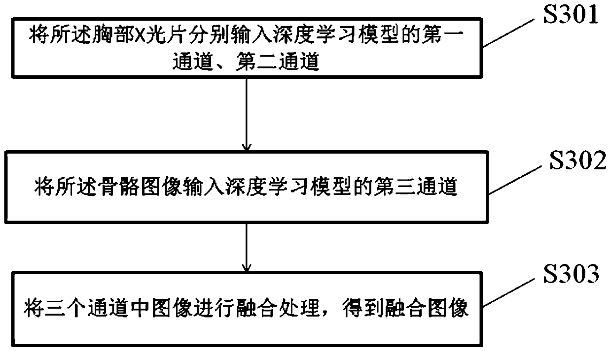
[0117] This embodiment is based on Embodiment 1. Such as image 3 As shown, the fusion processing of the chest X-ray film and the bone image to obtain the fusion image includes:

[0118] S301. Input the chest X-ray film into the first channel and the second channel of the deep learning model respectively;

[0119]S302. Input the bone image into the third channel of the deep learning model;

[0120] S303. Perform fusion processing on the images in the three channels to obtain a fusion image.

[0121] In a specific embodiment, such as Figure 4 As shown, the image screening model includes determining by the following method:

[0122] S401. Input a sample fusion image in the deep learning model;

[0123] S402. Use the classification network, detection network or segmentation network in the deep learning model to train and output the sample fusion image; for example, for scoliosis, which has a large abnormal area and obvious features, the abnormal bone image can be Use a cla...

Embodiment 3

[0144] Such as Figure 6 As shown, this embodiment discloses an image screening device, which includes:

[0145] Skeletal image acquisition module 601, used to acquire bone images from chest X-ray films;

[0146] An image fusion processing module 602, configured to perform fusion processing on the chest X-ray film and the bone image to obtain a fusion image;

[0147] The image screening module 603 is configured to input the fused image into an image screening model for testing, and obtain an image screening result; Learn to train certain models.

[0148] Further, as Figure 7 As shown, the skeleton image acquisition module 601 includes:

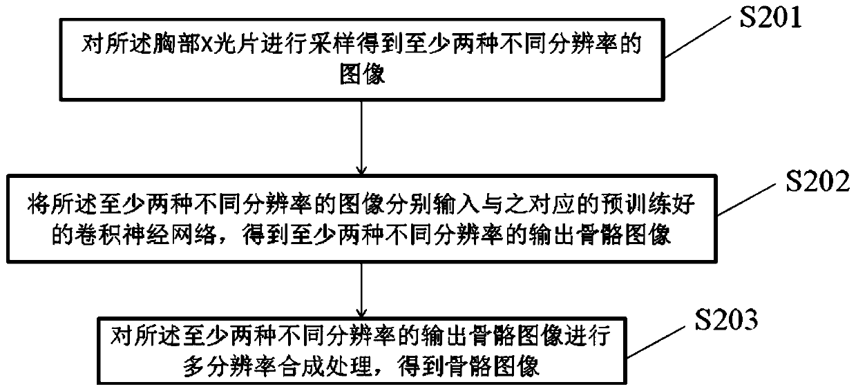
[0149] A resolution image acquisition module 6011, configured to sample the chest X-ray film to obtain at least two images with different resolutions;

[0150] The output bone image acquisition module 6012 is used to input the images of at least two different resolutions into the corresponding pre-trained convolutional neural network to ...


PUM


Abstract

The invention discloses an image screening method and device. The method comprises the steps of obtaining a bone image is acquired from a chest X-ray film; carrying out fusion processing on the chestX-ray film and the bone image to obtain a fusion image; inputting the fused image into an image screening model for testing to obtain an image screening result, wherein the image screening model comprises a model determined by deep learning training based on a sample fusion image and an abnormal skeleton tag of the sample fusion image. According to the method, the bone image generated by a similardual-energy subtraction technology can be generated on a conventional X-ray chest radiograph, so that the shielding of soft tissues on bones is greatly reduced, clearer and more detailed focus information is provided, the full-automatic screening of abnormal bone images is realized, and an image screening result with relatively high accuracy is obtained.

Description

technical field [0001] The invention relates to the field of image screening, in particular to an image screening method and device. Background technique [0002] Over the past few decades, chest radiography has been an important tool that has been widely used in clinical diagnosis. X-ray photography has become the preferred choice for chest examination because of its convenience, simplicity and economy. However, the diversity of diseases in chest radiographs and the limitations of 2D imaging in chest radiographs bring great challenges to doctors' diagnosis. [0003] Lodwick proposed a traditional machine learning method. He first extracted some quantitative features from the original chest radiograph, and then used these features to make a diagnosis of the disease by training a specific classifier. However, artificially selecting features is an extremely complicated matter, and there is no definite standard to judge which features are better. Deep learning avoids artific...

Claims


Application Information

Patent Timeline
IPC IPC(8): G06T7/00
Inventor 何志强郑介志
Owner SHANGHAI UNITED IMAGING INTELLIGENT MEDICAL TECH CO LTD
Why Eureka
Try Eureka