Fundus image retina blood vessel diameter automatic quantification method

A technology for retinal blood vessels and fundus images, which is applied in the field of medical image processing, can solve the problems of quantifying the size of blood vessels, difficulty in ensuring accuracy and consistency, poor repeatability, etc., and achieve the effect of reducing processing time

Active Publication Date: 2019-05-31
SHAOGUAN COLLEGE
View PDF9 Cites 7 Cited by
  • Summary
  • Abstract
  • Description
  • Claims
  • Application Information

AI Technical Summary

Problems solved by technology

Most of these methods are judged by manual observation, manual measurement and experience, and cannot technically accurately detect and extract retinal blood vessels and quantify the diameter of blood vessels, which will not be conducive to early prediction and auxiliary diagnosis of cardiovascular and cerebrova...

Method used


Image

  • Fundus image retina blood vessel diameter automatic quantification method
  • Fundus image retina blood vessel diameter automatic quantification method
  • Fundus image retina blood vessel diameter automatic quantification method

Examples


Embodiment Construction

[0083] In order to make the object, technical solution and advantages of the present invention more clear, the present invention will be further described in detail below in conjunction with the examples. It should be understood that the specific embodiments described here are only used to explain the present invention, not to limit the present invention.

[0084] Aiming at the problems of manual observation and manual measurement of blood vessel diameter in the prior art, labor intensity is high and time-consuming is long. The invention automatically measures the diameter of the retinal blood vessels through the image processing technology, provides early prediction and reference for auxiliary diagnosis for doctors, reduces the labor intensity of doctors, and improves diagnosis efficiency.

[0085] The application principle of the present invention will be described in detail below in conjunction with the accompanying drawings.

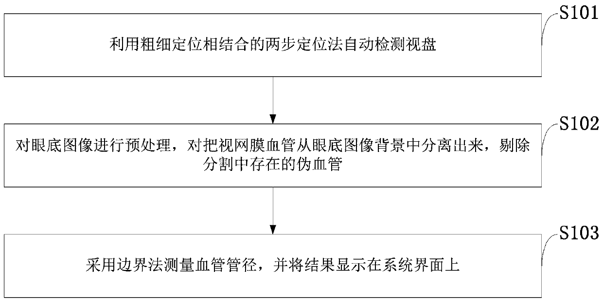
[0086] Such as figure 1 As shown, the method...


PUM


Abstract

The invention belongs to the technical field of medical image processing, and discloses a fundus image retina blood vessel diameter automatic quantification method. The method comprises the followingsteps of using a two-step positioning method combining coarse positioning and fine positioning to automatically detect the visual disc; preprocessing the fundus image, including denoising and enhancement processing of the fundus image; separating the retinal blood vessel from the background of the fundus image by adopting a multi-scale analysis method, and reconstructing and eliminating pseudo blood vessels in segmentation by utilizing mathematical morphology; and measuring the diameter of the blood vessel by using a boundary method, and displaying a result. On the basis of fundus image features, the fundus image feature extraction method firstly detects the optic disc, then enhances the image contrast ratio, segments the retinal blood vessel, finally measures the size of the retinal bloodvessel diameter, displays the result on the system interface, provides reference for early prediction and auxiliary diagnosis of clinicians, saves manpower and improves the efficiency.

Description

technical field [0001] The invention belongs to the technical field of medical image processing, and in particular relates to a method for automatically quantifying retinal vessel diameters in fundus images. Background technique [0002] At present, the commonly used existing technology in the industry is as follows: the fundus image of retinal blood vessels is an economical and effective clinical auxiliary method for ophthalmic diseases, and its development has attracted widespread attention. Changes in retinal arteriovenous intersections, arterial branches, venous paths, and morphology are important symptoms of retinal arteriosclerosis. When doctors observe fundus images, they mainly use radiography, projection, ophthalmoscopy, and calibration observation to measure the diameter of retinal vessels. Most of these methods are judged by manual observation, manual measurement and experience, and cannot technically accurately detect and extract retinal blood vessels and quanti...

Claims


Application Information

Patent Timeline
IPC IPC(8): G06T7/60G06T7/11G06T5/00
Inventor 罗忠亮
Owner SHAOGUAN COLLEGE
Why Eureka
Try Eureka