Method for discriminating lung tumor CT image by adopting high-dimensional feature selection

A CT image and feature selection technology, applied in the field of image processing, can solve the problems that the segmentation method cannot completely segment out lung tumors, low precision, and cannot detect a single shape feature

Inactive Publication Date: 2019-07-05
HARBIN UNIV OF SCI & TECH +1
View PDF4 Cites 7 Cited by
  • Summary
  • Abstract
  • Description
  • Claims
  • Application Information

AI Technical Summary

Problems solved by technology

In the image segmentation process, it is usually divided into threshold-based segmentation, edge-based segmentation, region-based segmentation and graph theory-based segmentation. A single segmentation method usually cannot completely segment the lung tumor in the image.
In the feature extraction process, due to the various shapes of lung tumors, it is impossible to detect them with a single sha

Method used


Image

  • Method for discriminating lung tumor CT image by adopting high-dimensional feature selection
  • Method for discriminating lung tumor CT image by adopting high-dimensional feature selection
  • Method for discriminating lung tumor CT image by adopting high-dimensional feature selection

Examples


Example Embodiment

[0035] The embodiments of the present invention will be described in detail below with reference to the accompanying drawings. It should be clear that the described embodiments are only a part of the embodiments of the present invention, rather than all the embodiments. Based on the embodiments of the present invention, all other embodiments obtained by those of ordinary skill in the art without creative work shall fall within the protection scope of the present invention.

[0036] Specific embodiments of the present invention are given below to help the understanding of the present invention.

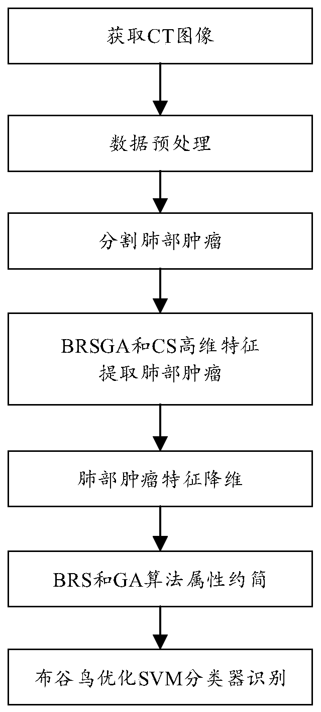
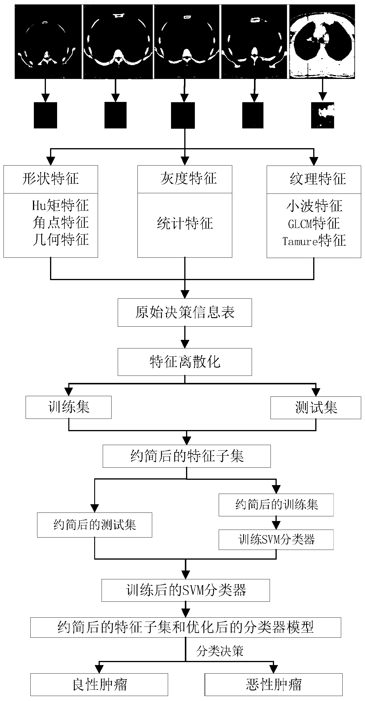
[0037] The present invention is based on the design of a threshold image segmentation model and a lung tumor classification model, and includes steps S101 to S103:

[0038] S101, image segmentation, the present invention uses the maximum between-class variance method (OTSU) to segment the preprocessed ROI region. Before image segmentation, the case image is preprocessed, and the sub-images ...


PUM


Abstract

The invention discloses a method for carrying out benign and malignant discrimination on a lung tumor CT image by adopting high-dimensional feature selection, and the method comprises the steps of obtaining a lung tumor CT image with a clear diagnosis conclusion in a lung image database, and forming a single sample; segmenting the preprocessed ROI by adopting a maximum between-cluster variance method so as to accurately describe the shape, texture and gray features of the lung tumor CT image; carrying out region feature extraction on the obtained lung CT image, and carrying out 82-dimensionalfeature component normalization on the extracted features so as to jointly quantify the ROI; carrying out attribute reduction by utilizing BRS and GA algorithms to reduce the time complexity and the space complexity of the classifier and improve the classification performance; and carrying out parameter global optimization and classification operation by using a cuckoo algorithm and an SVM mode, and carrying out lung tumor high-dimensional feature selection. The method has a very good auxiliary effect on lung cancer diagnosis, and the missed diagnosis rate and the misdiagnosis rate can be effectively reduced.

Description

technical field [0001] The present invention relates to the technical field of image processing, in particular to a method for distinguishing benign from malignant by selecting high-dimensional features of lung tumors based on CT images. Background technique [0002] With the development of computer-aided diagnosis research and the application of different modalities of medical image technology, the modalities are divided into pixel level, feature level and decision level according to different levels. The feature-level processing realizes the compression of the amount of information on the basis of retaining important information, and the processing speed is faster. In the process of image feature-level processing, the redundancy and correlation between features make the "curse of dimensionality" an NP-hard problem. Feature selection is an effective measure to solve this problem, which can effectively reduce the size of the feature space. dimensions to reduce time complexi...

Claims


Application Information

Patent Timeline
IPC IPC(8): G06T7/00G06T7/11G06T7/136G06K9/62
CPCG06T7/0012G06T7/11G06T7/136G06T2207/10081G06T2207/30096G06F18/2111G06F18/2411
Inventor 王进科赵聪聪祖宏亮黄飞杨博韬
Owner HARBIN UNIV OF SCI & TECH
Why Eureka
Try Eureka