
Method and system for generating standardized uptake value (SUV) based on medical image data

A medical imaging and data generation technology, applied in the field of medical imaging post-processing/analysis, can solve the problems that affect the speed of clinical case analysis, many human factors, and inaccuracy, so as to shorten the time for analyzing data, reduce inaccuracy, The effect of reducing the error rate

Pending Publication Date: 2019-07-16
XUANWU HOSPITAL OF CAPITAL UNIV OF MEDICAL SCI
View PDF7 Cites 1 Cited by
  • Summary
  • Abstract
  • Description
  • Claims
  • Application Information

AI Technical Summary

Problems solved by technology

However, the SUV algorithm in the post-processing workstation is relatively old, and studies have confirmed that the old SUV algorithm has obvious inaccuracies in liver edema
In addition, when the lesion area involves multi-layer images, workstation-based SUV analysis requires clinicians to manually record the SUV of each layer to calculate the average or maximum volume-based SUV, which seriously affects the speed of clinical case analysis, and human factors Larger errors lead to larger

Method used


Image

  • Method and system for generating standardized uptake value (SUV) based on medical image data
  • Method and system for generating standardized uptake value (SUV) based on medical image data
  • Method and system for generating standardized uptake value (SUV) based on medical image data

Examples


Embodiment Construction

[0020] The specific embodiments of the present invention are described below so that those skilled in the art can understand the present invention, but it should be clear that the present invention is not limited to the scope of the specific embodiments. For those of ordinary skill in the art, as long as various changes Within the spirit and scope of the present invention defined and determined by the appended claims, these changes are obvious, and all inventions and creations using the concept of the present invention are included in the protection list.

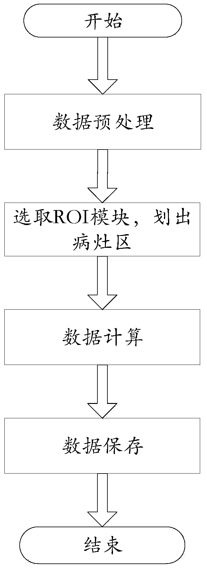
[0021] see attached image figure 1 , figure 1 A schematic flowchart illustrating a method for generating a standardized uptake value (SUV) based on medical image data is illustrated. The method comprises the steps of:

[0022] The first step: preprocessing the medical image data; wherein, the preprocessing includes converting the medical image data in DICOM format into NIFTI format, and also includes performing standard t...


PUM


Abstract

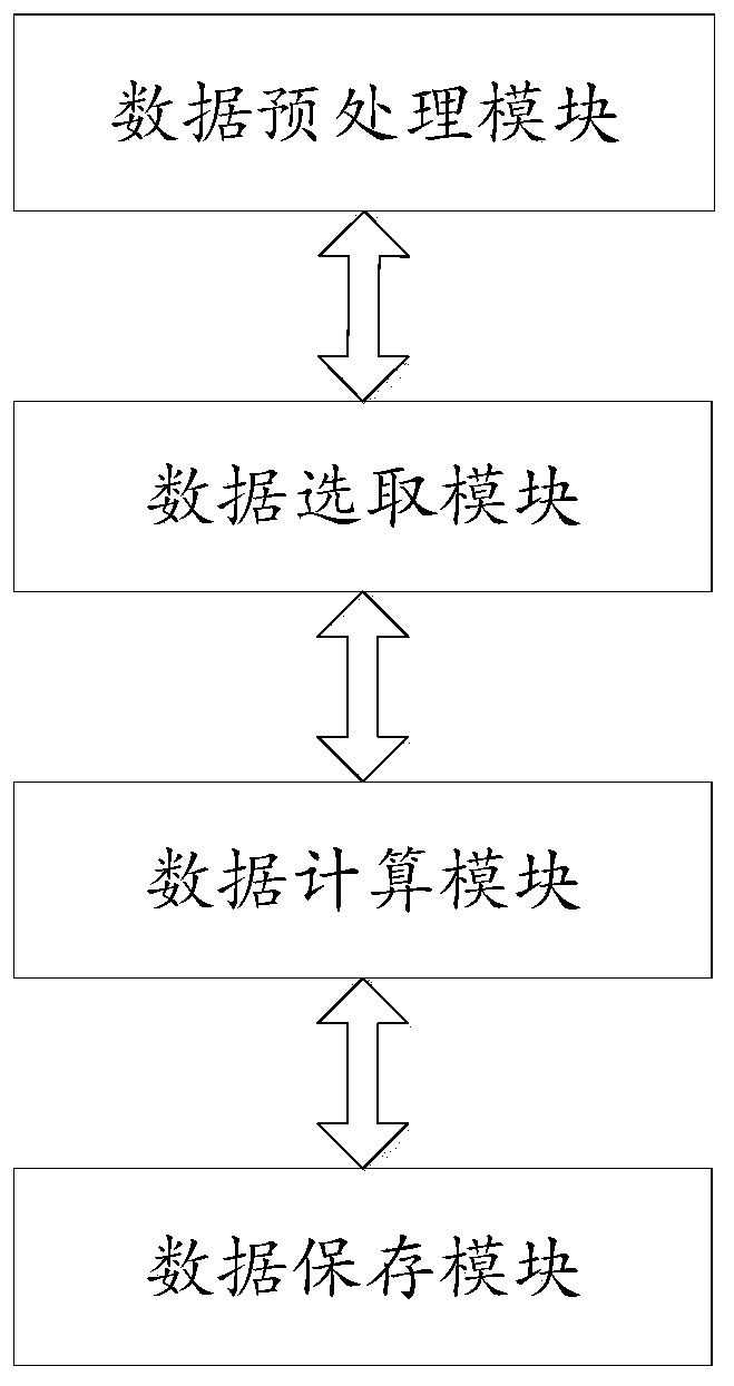
The present invention discloses a method for generating a standardized uptake value (SUV) based on medical image data. The method comprises the following steps: step one, the medical image data are preprocessed; step two, users select a module of region-of-interest for the preprocessed medical image data in the step one to draw a lesion region; step three, in the module of the region-of-interest,the standardized uptake value is calculated; and step four, the calculated results are subjected to data save. The system and method completely depend on data parameters stored by the system itself, do not require manual input by clinical staff, reduce inaccuracy caused by human factors, do not require the users to record and batch-save each data, save many cumbersome work, reduce an error rate, at the same time can manually divide the lesion region online, automatically complete and save all the required data results, are simple and rapid to operate, and greatly shorten time for clinical research doctors to analyze the data.

Description

technical field [0001] The invention relates to the technical field of post-processing / analysis of medical imaging, in particular to a method and system for generating a standardized uptake value (SUV) based on medical imaging data. Background technique [0002] Positron emission tomography (PET) is a medical imaging modality that allows the extraction of quantitative information related to the biodistribution of metabolically active contrast agents associated with FDG, FET, FLT, FMISO, etc. PET not only allows a visual representation of the distribution of the administered metabolically active contrast agent, but also allows quantification of how much radiopharmaceutical (also called radiotracer) has accumulated in a particular area. Using PET it is possible to determine exactly how much decay from radioisotopes has been counted in a given area. This makes it possible to compare these numbers with PET scans before or after, and to assess whether uptake and retention remain...

Claims


Application Information

Patent Timeline
IPC IPC(8): A61B6/00
CPCA61B6/5211
Inventor 卢洁赵国光殷雅彦
Owner XUANWU HOSPITAL OF CAPITAL UNIV OF MEDICAL SCI
Learn More