System and method for planning drainage of brain hematoma
A planning system and brain technology, applied in the planning system field of cerebral hematoma drainage, can solve the problems of secondary scanning, long-term planning, etc., and achieve the effect of fast puncture positioning, avoiding operation time, and avoiding blind puncture
- Summary
- Abstract
- Description
- Claims
- Application Information
AI Technical Summary
Problems solved by technology
Method used
Image
Examples
Embodiment 1
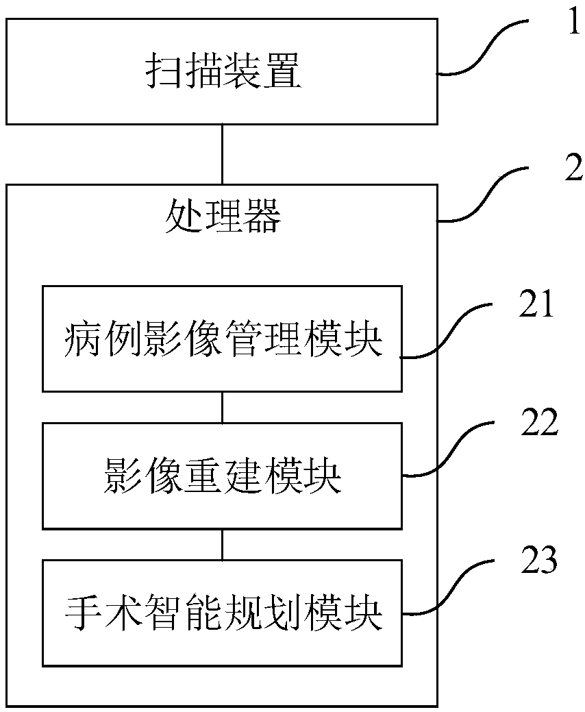
[0054] A planning system for brain hematoma drainage, such as figure 1 As shown, the planning system includes a scanning device 1 and a processor 2, and the processor 2 includes a case image management module 21, an image reconstruction module 22 and an intelligent operation planning module 23; the scanning device 1 includes CT, CTA, MRI At least one of , MRA and DSA.
[0055] The scanning device 1 is used to scan the patient once before the operation to obtain scanning image data and send it to the case image management module 21;
[0056] The case image management module 21 is used to receive and store the scanned image data;
[0057]The image reconstruction module 22 is used to extract the scanned image data from the case image management module 21, generate a three-dimensional simulated image of the patient according to the scanned image data and send it to the intelligent operation planning module 23;
[0058] The intelligent operation planning module 23 is used to gene...
Embodiment 2
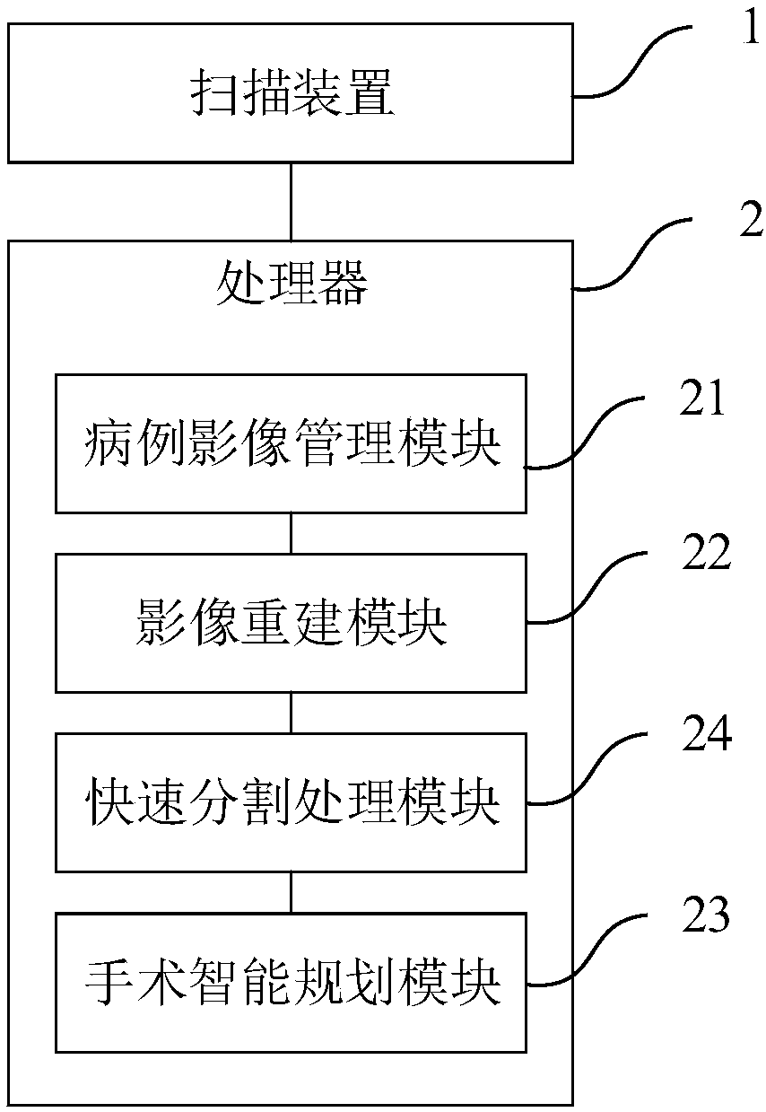
[0061] Such as figure 2 As shown, the planning system for cerebral hematoma drainage in this embodiment is further improved on the basis of Embodiment 1, and the processor 2 also includes a rapid segmentation processing module 24;
[0062] The rapid segmentation processing module 24 is used to extract the scanned image data from the case image management module 21, and extract craniocerebral hematoma data from the scanned image data according to the gray value area growth algorithm, and the craniocerebral hematoma Data include cerebral hematoma location and cerebral hematoma size;
[0063] The intelligent operation planning module 23 is used to generate the optimal puncture path according to the brain hematoma data and the three-dimensional simulation image.
[0064] In this embodiment, the tomographic image data obtained by scanning, according to the gray value information of the data, based on the algorithm principle of gray value area growth, can obtain the target point a...
Embodiment 3
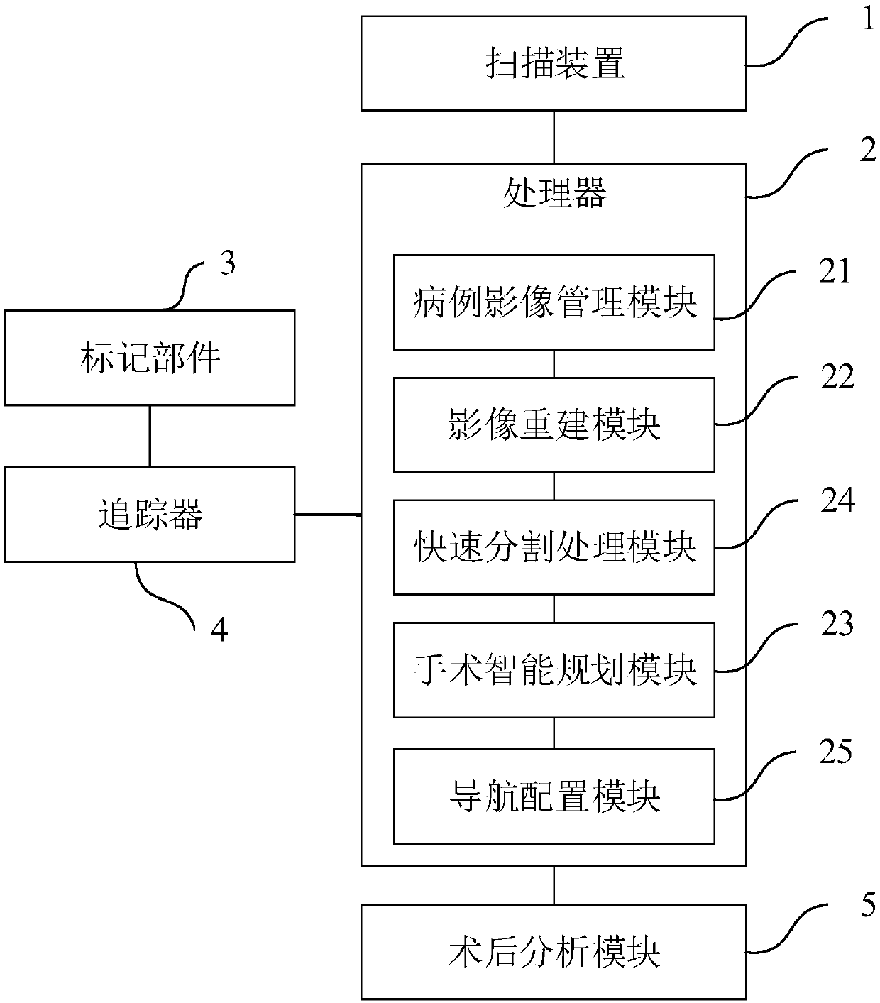
[0066] Such as image 3 As shown, the planning system for cerebral hematoma drainage in this embodiment is further improved on the basis of Embodiment 1, and the planning system also includes a tracker 4 and a marking part 3, and the marking part 3 is set at the patient's surgical site. On the body and surgical instruments within a certain preset range, the processor 2 also includes a navigation configuration module 25;
[0067] The scanning device 1 is used to obtain scanned image data including the image of the marking part 3 before the operation;
[0068] The tracker 4 is used to obtain the static data of the marking part 3 set on the patient's body during the operation and send it to the navigation configuration module 25, and is also used to obtain the real-time data set on the surgical instrument during the operation. Mark the dynamic data of component 3 and send to the navigation configuration module 25;
[0069] The navigation configuration module 25 is used to extra...
PUM
 Abstract
Description
Claims
Application Information
 - R&D Engineer
- R&D Manager
- IP Professional
- Industry Leading Data Capabilities
- Powerful AI technology
- Patent DNA Extraction
Browse by: Latest US Patents, China's latest patents, Technical Efficacy Thesaurus, Application Domain, Technology Topic, Popular Technical Reports.
© 2024 PatSnap. All rights reserved.Legal|Privacy policy|Modern Slavery Act Transparency Statement|Sitemap|About US| Contact US: help@patsnap.com


