Image feature acquisition method of prostate MRI three-dimensional image

An image feature, three-dimensional image technology, applied in image analysis, medical image, image data processing and other directions, can solve the problems of increasing the cost and cycle of diagnosis, and cannot be automatically segmented, so as to improve the efficiency and accuracy of diagnosis and reduce interference.

Pending Publication Date: 2019-08-30
WONDERS INFORMATION
View PDF8 Cites 12 Cited by
  • Summary
  • Abstract
  • Description
  • Claims
  • Application Information

AI Technical Summary

Problems solved by technology

[0004] At present, there are some methods for prostate cancer discrimination, and some use deep learning methods to use the entire image image as the input of the model, but some irrelevant tissues around the prostate organ will be included in the MRI scanning process, and this method will vary due to the surrounding organs Differences in non-prostate regions lead to misjudgment; other methods only use T2WI images, ignoring the characteristics of other seq

Method used


Image

  • Image feature acquisition method of prostate MRI three-dimensional image
  • Image feature acquisition method of prostate MRI three-dimensional image
  • Image feature acquisition method of prostate MRI three-dimensional image

Examples


Embodiment Construction

[0031] Below in conjunction with specific embodiment, further illustrate the present invention. It should be understood that these examples are only used to illustrate the present invention and are not intended to limit the scope of the present invention. In addition, it should be understood that after reading the teachings of the present invention, those skilled in the art can make various changes or modifications to the present invention, and these equivalent forms also fall within the scope defined by the appended claims of the present application.

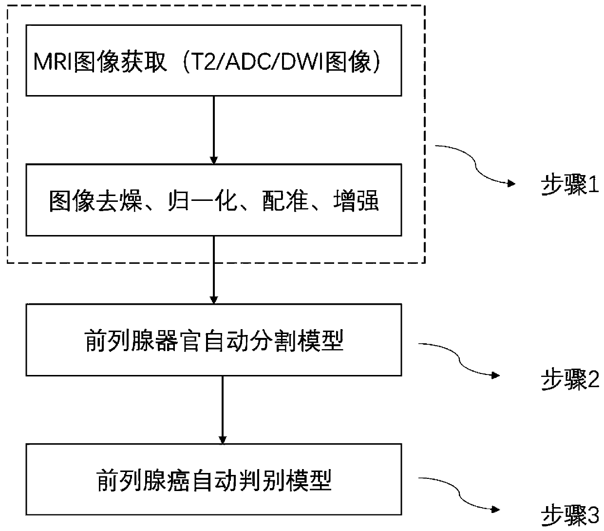
[0032] In this embodiment, applying the image feature acquisition method of a prostate MRI three-dimensional image provided by the present invention to the automatic discrimination of prostate cancer includes the following steps:

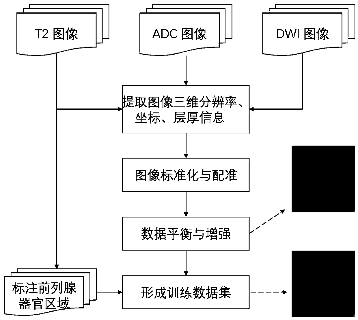
[0033] 1. Acquire and preprocess the image, the process is as follows figure 2 As shown, it mainly includes the following steps:

[0034] 101) Data collection: Collect prostate MRI image data sets...


PUM


Abstract

The invention relates to an image feature acquisition method of a prostate MRI three-dimensional image. The method comprises: the prostate T2WI image being subjected to automatic organ segmentation; and mapping the prostate organ region to the registered ADC and DWI image based on a segmentation result to obtain a multi-parameter prostate organ region serving as input of a discrimination model, and obtaining image characteristics by combining the multi-parameter MRI image and a deep learning algorithm. The method is based on a large amount of prostate image data, the prostate organ automatic segmentation model is established, the interference of irrelevant background information on the discrimination model is reduced, the characteristics of the multi-parameter MRI image are fused by usinga deep learning method, high-precision prostate cancer characteristic extraction is realized, and a basis is provided for improving the prostate cancer diagnosis efficiency and accuracy.

Description

technical field [0001] The invention relates to an image feature acquisition method of a prostate MRI three-dimensional image. The acquired image features can be used for auxiliary diagnosis of prostate cancer, and belong to the technical field of computer-aided diagnosis of medical images. Background technique [0002] Prostate cancer is a common malignant tumor in men and the second most common cancer in men worldwide. The incidence of prostate cancer in Chinese men is also increasing day by day. Magnetic resonance imaging (MRI) examination has become an important method for the diagnosis of prostate cancer in hospitals due to its advantages of non-invasive examination, diversified scanning images, and prominent focus signals. MRI imaging includes T2WI (T2-weighted imaging), DWI (diffusion-weighted imaging), ADC ( Surface diffusion coefficient) and other sequences assist radiologists in diagnosis. Among them, the T2WI image provides a clearer tissue contrast, and the DWI ...

Claims


Application Information

Patent Timeline
IPC IPC(8): G06K9/62G06N3/04G06T7/11G16H30/40
CPCG16H30/40G06T7/11G06N3/045G06F18/214Y02T10/40
Inventor 张敬谊王培军丁偕王伟王瑜王磊曹学香
Owner WONDERS INFORMATION
Why Eureka
Try Eureka