
Brain glioma region automatic segmentation method

A technology for automatic segmentation of glioma, applied in the field of medical image analysis, can solve the problems of overall model redundancy, low segmentation accuracy, and dependence, and achieve the effects of reducing interference, ensuring segmentation effect, and reducing model size

Active Publication Date: 2019-08-30
UNIV OF SCI & TECH OF CHINA
View PDF6 Cites 9 Cited by
  • Summary
  • Abstract
  • Description
  • Claims
  • Application Information

AI Technical Summary

Problems solved by technology

[0003] At present, the existing research methods often use cascaded neural networks to deal with the segmentation of different regions, which makes the overall model very redundant, and the training of the networks is independent of each other, while the network input of the latter stage is heavily dependent on the previous stage. network, therefore, the final segmentation accuracy is also lower

Method used


Image

  • Brain glioma region automatic segmentation method
  • Brain glioma region automatic segmentation method
  • Brain glioma region automatic segmentation method

Examples


Embodiment Construction

[0011] The technical solutions in the embodiments of the present invention will be clearly and completely described below in conjunction with the accompanying drawings in the embodiments of the present invention. Obviously, the described embodiments are only some of the embodiments of the present invention, not all of them. Based on the embodiments of the present invention, all other embodiments obtained by persons of ordinary skill in the art without making creative efforts belong to the protection scope of the present invention.

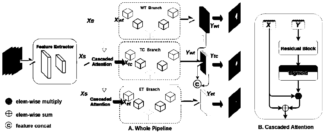
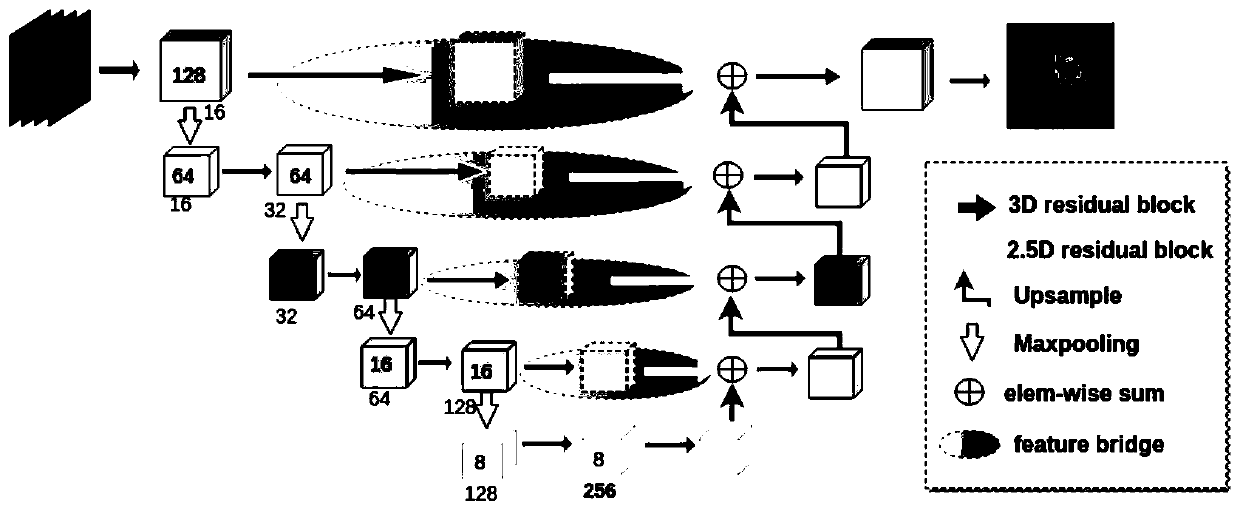
[0012] The embodiment of the present invention provides a method for automatic segmentation of brain glioma regions, the main process is as follows: use the segmentation network of the cascade attention mechanism to process the multimodal three-dimensional brain volume data MRI images, and the cascade attention mechanism The front-end of the segmentation network is a shared feature extractor, and its back-end is connected to three codec branches wit...


PUM


Abstract

The invention discloses an automatic segmentation method for a brain glioma region. The method comprises the steps that a multi-mode three-dimensional brain body data MRI image is processed through asegmentation network of a cascade attention mechanism, the front end of the segmentation network of the cascade attention mechanism is a shared feature extractor, and the rear end of the segmentationnetwork of the cascade attention mechanism is connected with three coding and decoding device branches of the same structure; the feature extractor respectively inputs the extracted feature map into three codec branches, and the three codec branches are used for segmenting different areas; an attention mechanism module is arranged between every two adjacent encoder and decoder branches and used for capturing the spatial hierarchical relationship of different areas, and finally different areas in the brain glioma area are segmented through the three encoder and decoder branches. By means of themethod, automatic segmentation of the brain glioma area can be rapidly and accurately achieved.

Description

technical field [0001] The invention relates to the technical field of medical image analysis, in particular to a method for automatically segmenting brain glioma regions. Background technique [0002] Due to the heterogeneity and spatial diversity of gliomas, it is of great significance to accurately segment tumor regions automatically. [0003] At present, the existing research methods often use cascaded neural networks to deal with the segmentation of different regions, which makes the overall model very redundant, and the training of the networks is independent of each other, while the network input of the latter stage is heavily dependent on the previous stage. network, therefore, the final segmentation accuracy is also lower. Contents of the invention [0004] The object of the present invention is to provide a method for automatically segmenting brain glioma regions, which can quickly and accurately realize automatic brain glioma region segmentation. [0005] The ...

Claims


Application Information

Patent Timeline
Patent Type & Authority Applications(China)
IPC IPC(8): G06T7/11
CPCG06T7/11G06T2207/10088G06T2207/20081G06T2207/30016G06T2207/30096
Inventor 张勇东徐海谢洪涛
Owner UNIV OF SCI & TECH OF CHINA
Learn More