X-ray imaging analysis method and device based on AI and readable storage medium

An analysis device and analysis method technology, which is applied in the fields of radiodiagnostic instruments, radiodiagnostic equipment control, medical science, etc., can solve problems such as easy fatigue, reduce errors and missed detections, lower technical requirements, and lower technical thresholds Effect

Pending Publication Date: 2019-10-22
CARERAY DIGITAL MEDICAL TECH CO LTD
View PDF4 Cites 5 Cited by
  • Summary
  • Abstract
  • Description
  • Claims
  • Application Information

AI Technical Summary

Problems solved by technology
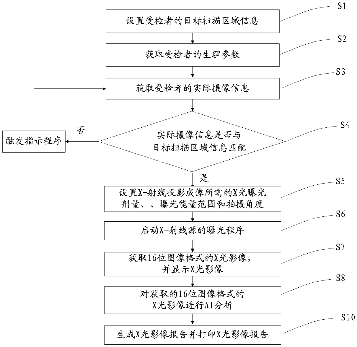
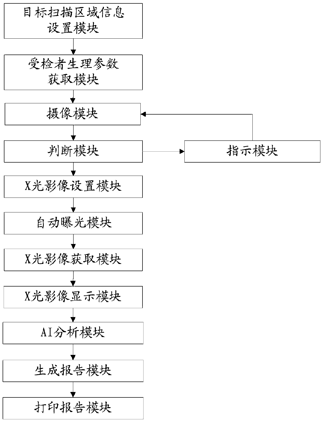
The work is highly repetitive and monotonous, and gets tired easily

Method used


Image

  • X-ray imaging analysis method and device based on AI and readable storage medium
  • X-ray imaging analysis method and device based on AI and readable storage medium
  • X-ray imaging analysis method and device based on AI and readable storage medium

Examples


Embodiment Construction

[0053] In order to enable those skilled in the art to better understand the solutions of the present invention, the following will clearly and completely describe the technical solutions in the embodiments of the present invention in conjunction with the drawings in the embodiments of the present invention. Obviously, the described embodiments are only It is an embodiment of a part of the present invention, but not all embodiments. Based on the embodiments of the present invention, all other embodiments obtained by persons of ordinary skill in the art without making creative efforts shall fall within the protection scope of the present invention.

[0054] It should be noted that the terms "first" and "second" in the description and claims of the present invention and the above drawings are used to distinguish similar objects, but not necessarily used to describe a specific sequence or sequence. It is to be understood that the data so used are interchangeable under appropriate ...


PUM


Abstract

The invention discloses an X-ray imaging analysis method and device based on AI and a readable storage medium. The analysis method comprises the steps that target scanning area information of an examinee is set, wherein the target scanning area information comprises a to-be-scanned target area; physiological parameters of the examinee are acquired; according to the set target scanning area information and the acquired physiological parameters of the examinee, actual camera shooting information of the examinee is acquired; whether or not the actual camera shooting information is matched with the target scanning area information is judged; the X-ray exposure hose, exposure energy range and shooting angle required by X-ray projection imaging are set; an exposure procedure of an X-ray source is started; an X-ray image of a 16-bit image form is acquired and displayed; the acquired X-ray image of the 16-bit image form is subjected to AI analysis. According to the analysis device, the technical threshold of a user is greatly lowered, the examination efficiency is effectively improved, and the fault and missing examination of diagnosis are reduced.

Description

technical field [0001] The invention relates to the field of X-ray projection imaging equipment, in particular to an AI-based X-ray imaging analysis method, device and readable storage medium. Background technique [0002] In the current X-ray projection imaging equipment industry, software development and image processing are largely limited by the division of labor of technicians or physicians, as well as the existing environmental conditions. For example, the technician responsible for taking X-rays needs to tell the patient how to position correctly and set the shooting dose according to the visual observation of the patient's condition. Work is highly repetitive, monotonous, and tires easily. Patients often need to rush between the consulting room and the billing office and the imaging department. The technicians in the imaging department are basically only responsible for the shooting of X-ray images, but not involved in the diagnosis; while the physicians are respon...

Claims


Application Information

Patent Timeline
Patent Type & Authority Applications(China)
IPC IPC(8): A61B6/00
CPCA61B6/4411A61B6/54A61B6/544A61B6/545A61B6/563A61B6/566A61B6/467
Inventor 赵杰凌骏
Owner CARERAY DIGITAL MEDICAL TECH CO LTD
Why Eureka
Try Eureka