
Internally implanted anatomical elbow joint stabilizer

An anatomical, elbow-joint technology, applied in the field of medical devices, can solve problems such as affecting the appearance and wearing clothes, making patients uncomfortable, and easily damaging articular cartilage, so as to reduce the risk of skin necrosis, have little impact on normal life, and reduce wound infection. Effect

Pending Publication Date: 2019-12-17
XIANGYA HOSPITAL CENT SOUTH UNIV
View PDF1 Cites 2 Cited by
  • Summary
  • Abstract
  • Description
  • Claims
  • Application Information

AI Technical Summary

Problems solved by technology

However, the direct use of Kirschner wires to fix the elbow joint is easy to damage the articular cartilage, and the infection of the needle tract will lead to septic arthritis, and the natural contraction force of the elbow muscles will give a large reaction force to the steel needle used for fixation, often resulting in elbow joint fixation Weakness and Kirschner wire breakage; plaster splints and braces also cannot provide sufficient stability, especially for obese patients, and the fixation effect is not good; more importantly, the above methods all fix the elbow joint in a static state , the elbow joint cannot perform flexion and extension activities, and prolonged immobilization may cause the patient's elbow joint to become stiff again, and the flexion and extension activities are limited
Although the articulated external fixator can not only stabilize the elbow joint to prevent dislocation, but also ensure the flexion and extension of the elbow joint during the postoperative rehabilitation stage, the external fixator is bulky and heavy, and placing it on the upper arm will affect the appearance and wear clothes. Due to the effect of gravity , the patient is more uncomfortable, and there is also a high incidence of needle pain, infection and loosening, and the risk of damage to the radial nerve during screw placement

Method used


Image

  • Internally implanted anatomical elbow joint stabilizer
  • Internally implanted anatomical elbow joint stabilizer
  • Internally implanted anatomical elbow joint stabilizer

Examples


Embodiment Construction

[0018] The present invention will be further described in detail below in conjunction with the accompanying drawings and embodiments. In order to more clearly illustrate the technical solutions in the embodiments of this patent or the prior art, the accompanying drawings that need to be used in the description of the embodiments or the prior art will be described below. Brief introduction, it is obvious that the drawings in the following description are only some embodiments of the present invention, and those skilled in the art can also obtain other drawings according to these drawings without creative work .

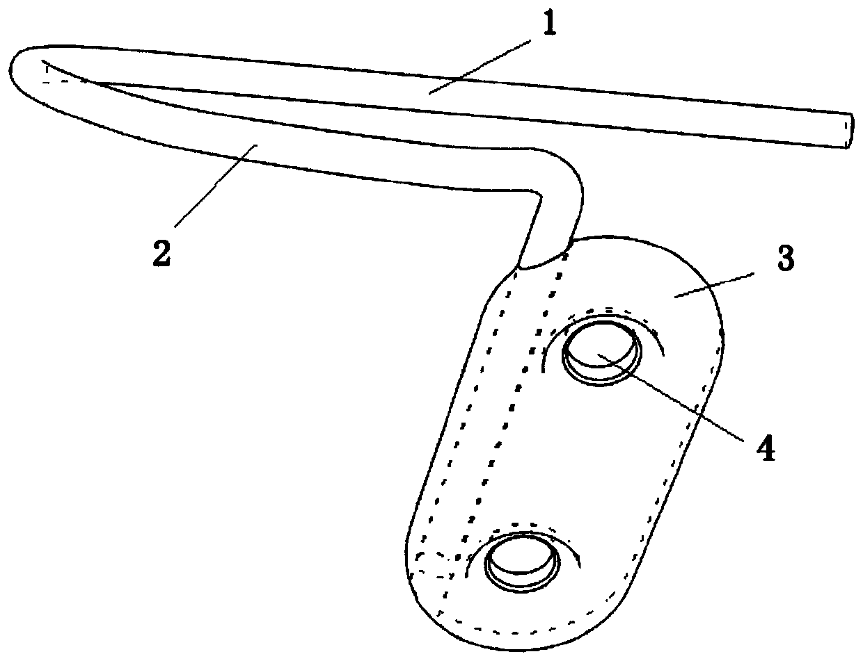
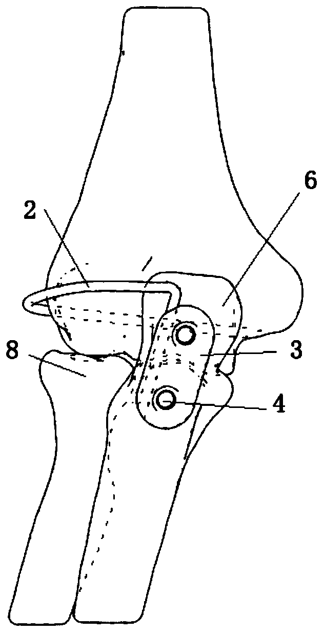
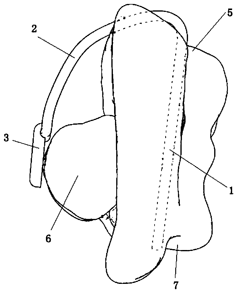
[0019] Such as Figures 1 to 6 As shown, the implantable anatomical elbow joint stabilizer includes a crossbar 1 that is placed in the rotation axis of the humerus capitellum 5 as the rotation axis of the humerus, a fixed plate 3 that is fixed on the olecranon 6 by screws, and the The connecting rod 2 connected to the cross bar 1 and the fixed plate 3 has a distance b...


PUM


Abstract

The invention discloses an internally implanted anatomical elbow joint stabilizer. The internally implanted anatomical elbow joint stabilizer comprises a transverse rod placed in the rotating axis ofa capitulum humerus to serve as a humerus rotating shaft, a fixing plate fixed to ulna olecranon through screws, and a connecting rod connecting the transverse rod with the fixing plate, spacing is formed between the connecting rod and an ulna, between the connecting rod and a radius as well as between the connecting rod and a humerus, the spacing enables the connecting rod not to interfere with the ulna, the radius and the humerus during bending and stretching movement, and the transverse rod, the connecting rod, the fixing plate and the screws are all made of degradable implanting materials.According to the internally implanted anatomical elbow joint stabilizer, after an operation, bending and stretching movement can be achieved, the stability of an elbow joint can be maintained under bending and stretching movement, the appearance of the elbow and clothes wearing are not affected after the operation, stent-tract infection is avoided, the risks of wound infection and skin necrosis are reduced, and a secondary operation for taking out the stabilizer is not needed.

Description

technical field [0001] The invention relates to the field of medical devices, in particular to an implanted anatomical elbow stabilizer. Background technique [0002] Elbow instability caused by injury or release surgery is difficult to manage in orthopedic practice. If the elbow joint is unstable and is not fixed, it will affect the upper limb function of the patient; if it is not handled properly, it will easily lead to secondary or multiple operations, adding additional pain to the patient. The following situations are examples of complex elbow instability: 1. Elbow triad injury and unstable coronoid fracture; 2. Elbow dislocation with severe soft tissue injury; After thorough release of soft tissue and / or excision of heterotopic ossification. The above-mentioned complex elbow joint injuries may still be unstable after bony structure fixation and ligament repair, and the existing traditional treatment methods for elbow joint instability include: 1. Directly nail the elb...

Claims


Application Information

Patent Timeline
Patent Type & Authority Applications(China)
IPC IPC(8): A61B17/80
CPCA61B17/8061A61B17/808A61B17/809
Inventor 成亮朱勇林涨源王小鹏
Owner XIANGYA HOSPITAL CENT SOUTH UNIV
Learn More