Retinal vessel segmentation method and system based on retinal fundus image

A retinal blood vessel and fundus image technology, applied in the field of image processing, can solve the problems of long test time, slow convergence speed, slowness, etc., to solve the data imbalance and speed up the training process.

Active Publication Date: 2020-01-14
BEIHANG UNIV +1
View PDF4 Cites 8 Cited by
  • Summary
  • Abstract
  • Description
  • Claims
  • Application Information

AI Technical Summary

Problems solved by technology

However, these patch-based methods have slow convergence speed, long testing time, and cannot obtain real-time results, so they are less applicable in clinical applications
For small data sets, previous methods have adopted a variety of data enhancement methods, which apply spatial adaptive contrast enhancement technology to retinal fundus images for blood vessel segmentation, and use static wavelet transform (SWT) to perform retinal fundus image segmentation. preprocessing, but SWT preprocessing is complicated and slow

Method used


Image

  • Retinal vessel segmentation method and system based on retinal fundus image
  • Retinal vessel segmentation method and system based on retinal fundus image
  • Retinal vessel segmentation method and system based on retinal fundus image

Examples


Embodiment Construction

[0022] In order to further explain the features of the present invention, please refer to the following detailed description and drawings of the present invention. The attached drawings are for reference and explanation purposes only, and are not used to limit the protection scope of the present invention.

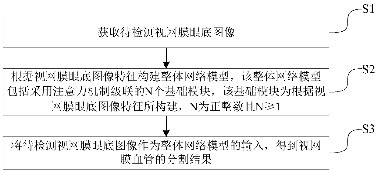
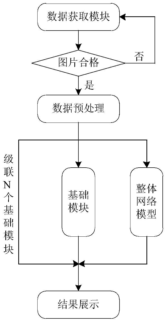
[0023] Such as Figure 1-Figure 2 As shown, this embodiment discloses a retinal blood vessel segmentation method based on retinal fundus images, which includes the following steps S1 to S3:

[0024] S1. Obtain a fundus image of the retina to be detected;

[0025] S2. Construct an overall network model based on the features of the retinal fundus image, the overall network model includes N basic modules cascaded using an attention mechanism, the basic module is constructed based on the features of the retinal fundus image, N is a positive integer and N≥1;

[0026] S3. Use the retinal fundus image to be detected as the input of the overall network model to obtain the segmentation re...


PUM


Abstract

The invention discloses a retinal vessel segmentation method and system based on a retinal fundus image, and belongs to the technical field of image processing, and the method comprises the steps: obtaining a to-be-detected retinal fundus image; constructing a basic module according to the retinal fundus image features; cascading N basic modules to serve as a final network model, the to-be-detected retinal fundus image serving as input of the whole network model, and obtaining a segmentation result of retinal vessels. The foreground characteristics of the previous basic module and the originalpicture are transmitted to the next basic module together, so that the rear basic module can inherit the learning experience of the front basic module, the training process is accelerated, and the problem of data imbalance is effectively solved; the to-be-detected retinal fundus image is used as the input of the overall model S-UNet, and the obtained segmentation result of the retinal blood vessel is more accurate.

Description

Technical field [0001] The present invention relates to the technical field of image processing, in particular to a method and system for retinal blood vessel segmentation based on retinal fundus images. Background technique [0002] Ophthalmologists usually check the retinal fundus images to assess the clinical condition of retinal blood vessels, which are important indicators for diagnosing various eye diseases. However, manual marking of retinal blood vessels in these images is time-consuming and cumbersome, and requires rich clinical experience. Therefore, real-time automatic segmentation of retinal blood vessels is very necessary, and it has also attracted much attention in recent years. [0003] Existing retinal vessel segmentation methods can be divided into unsupervised and supervised methods. For unsupervised methods, it is necessary to manually design feature extraction rules based on given data samples, so as to distinguish between blood vessels and background tissues b...

Claims


Application Information

Patent Timeline
Patent Type & Authority Applications(China)
IPC IPC(8): G06T7/00G06T7/10G06N3/04
CPCG06T7/0012G06T7/10G06T2207/30041G06T2207/30101G06T2207/20081G06N3/045
Inventor 张冀聪王华胡静斐
Owner BEIHANG UNIV
Why Eureka
Try Eureka