
Motion control method for flexible endoscope operation robot

A technology of motion control and endoscopy, applied in the field of medical robots, can solve the problems of inconvenient operation of endoscopes by robots

Inactive Publication Date: 2020-02-04
NANKAI UNIV
View PDF0 Cites 7 Cited by
  • Summary
  • Abstract
  • Description
  • Claims
  • Application Information

AI Technical Summary

Problems solved by technology

However, the traditional respiratory motion model constructed based on ultrasound images can only reflect the general law of respiratory motion, and cannot directly track organs or specific targets, it is difficult to establish personalized respiratory motion models for different human bodies; or the traditional method needs to arrange additional The sensor is used to collect displacement information, which may cause inconvenience to the robot operating the endoscope; there is no effective method to compensate the movement of the robot operating the endoscope according to the breathing motion model of human organs

Method used


Image

  • Motion control method for flexible endoscope operation robot
  • Motion control method for flexible endoscope operation robot
  • Motion control method for flexible endoscope operation robot

Examples


Embodiment 1

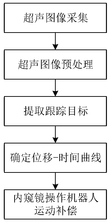
[0030] The invention proposes a motion control method for a flexible endoscope operating robot, which is used to eliminate the displacement deviation between the organ and the manipulator at the end of the endoscope caused by respiratory movement after the endoscope enters the human cavity.

[0031] [1] Firstly, before the endoscope enters the human cavity and after all preparations are completed, the ultrasonic images of the organs under the condition of stable human breathing are collected.

[0032] [2] Do filter preprocessing on the collected ultrasound images.

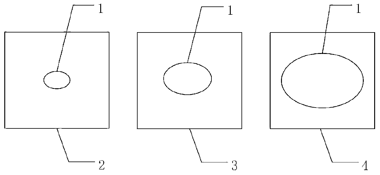
[0033] [3] Extract the outline of the target organ or the key feature points in the tissue and organ from the ultrasound image.

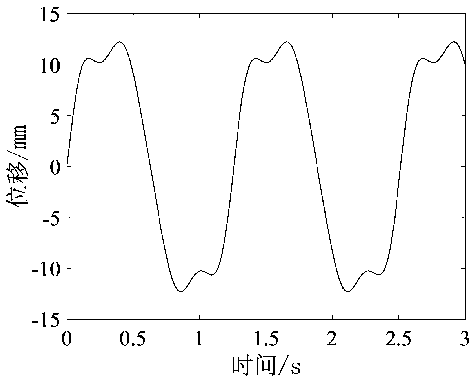
[0034] [4] Then record the position of the center point or feature point of the organ contour at different times, so as to determine the displacement-time curve of the human organ in the up and down direction, such as image 3 .

[0035] [5] If Figure 4 As shown, after the endoscope ...


PUM


Abstract

The invention relates to a motion control method for a flexible endoscope operation robot. The method comprises the steps of firstly, obtaining image information within a complete respiratory motion cycle of a human body; accurately determining the edge contour of organ tissue by using a medical image wave filtering and partitioning algorithm; determining displacement change condition of the organs along with the time according to the edge contour information, and drawing a displacement-time curve under respiration; and under the condition that the displacement-time curve of the organ is determined, performing motion compensation on the endoscope operation robot by referencing the cycle of the displacement-time curve and a fluctuation amplitude. A real-time ultrasound image is used for directly tracking the motion of the organs, so that the respiratory motion law of different human bodies and the same human body in different time ranges can be directly displayed; the displacement-timecurve of the organs under the breathing state, obtained by collected ultrasonic images and orifical hole images acquired by the endoscope are registered, so that compensation can be performed on displacement deviation between the endoscope and human organs generated by breathing.

Description

technical field [0001] The invention belongs to the technical field of medical robots, and relates to a motion compensation method for a medical robot under the action of respiratory motion when an endoscope enters a human body. Background technique [0002] In recent years, minimally invasive surgical robots represented by flexible endoscopes have played a huge role in practical applications, with the advantages of less trauma to the human body, fast postoperative recovery, and repeatable operations. [0003] Before the flexible endoscope enters the human cavity, the corresponding three-dimensional model is first established by collecting ultrasonic images of the human body to determine the target position and plan the path, and then cooperate with the navigation system endoscope operating robot to guide the end manipulator to the target position. [0004] At present, the three-dimensional models constructed based on CT images are generally static, and the size and shape of...

Claims


Application Information

Patent Timeline
Patent Type & Authority Applications(China)
IPC IPC(8): A61B34/30A61B34/10A61B34/00A61B34/20
CPCA61B34/20A61B34/30A61B34/70A61B2034/105A61B2034/301
Inventor 代煜陈通张建勋
Owner NANKAI UNIV
Learn More