
Blind rivet for adapting biological tissue and device for setting the same, in particular through the instrument channel of an endoscope

a blind rivet and biological tissue technology, applied in the field of self-piercing blind rivets, can solve the problems of inconvenient suturing, difficult endoscopy use, and additional instruments, and achieve the effects of reducing the risk of infection, and reducing the safety of surgical instruments

Inactive Publication Date: 2014-03-04
TECH UNIV MUNCHEN
View PDF37 Cites 23 Cited by
  • Summary
  • Abstract
  • Description
  • Claims
  • Application Information

AI Technical Summary

Benefits of technology

The invention provides a connecting element (a blind rivet) and a device for setting such elements, which can connect any number of tissue layers of any thickness with a contacting force. This means that surgeons can choose a rivet that can handle their specific needs. The blind rivet has a carrier with a fixed clip at the distal end, which pierces the tissue, and a holding device at the proximal side of the carrier to keep the rivet in place. The holding device can be a clip or a different device, like a medication dispenser or a diagnosis device with telemetric transfer. This invention can be used in the instrument channel of a flexible endoscope. Overall, the invention simplifies the process of connecting tissue layers and allows for greater precision and control during surgery.

Problems solved by technology

Their applications are limited, however, mainly to fixing implants or tissue to bones, as described in U.S. Pat. No. 6,241,732 B1 or the WO 99 / 62418.
This rivet, however, mainly is not constructed as a blind rivet and must instead be serviced from both sides, which makes use in endoscopy difficult and requires an additional instrument.
However, since these grippers do not attain even approximately the freedom of movement and the movement possibilities of the hand, this suturing is very difficult, uncertain and time consuming.
However, even with such a device, the most serious disadvantage of the suturing technique, namely, the knotting of the suture, cannot be eliminated.
Accordingly, because of their complexity and the therefrom resulting overall size, it has not yet been possible to insert such devices into the instrument channels of flexible endoscopes.
Once again, however, it is necessary to use a further instrument, so that the instrument channel of an endoscope cannot be used by itself.
It is therefore not possible to connect tissues of different or unknown thicknesses using one and the same clip.
This represents a major limitation for the surgeon.
Associated with this is the fact that the contacting force, with which the tissue is held together, is also not adjustable, so that the surgeon cannot gain any feeling for the quality of the connection.
Moreover, the functional reliability of the clip is also a cause for concern, since a stop has not been provided for the proximal fixation element, so that it may happen that the complete clip can be pushed through the tissue and, as it were, fall out on the other side.
It is also not possible here to load the clips into a magazine, that is, to set several clips consecutively with one instrument.

Method used


Image

  • Blind rivet for adapting biological tissue and device for setting the same, in particular through the instrument channel of an endoscope
  • Blind rivet for adapting biological tissue and device for setting the same, in particular through the instrument channel of an endoscope
  • Blind rivet for adapting biological tissue and device for setting the same, in particular through the instrument channel of an endoscope

Examples


Embodiment Construction

[0023]The preferred embodiments of the present invention will now be described with reference to FIGS. 1-11 of the drawings. Identical elements in the various figures are designated with the same reference numerals.

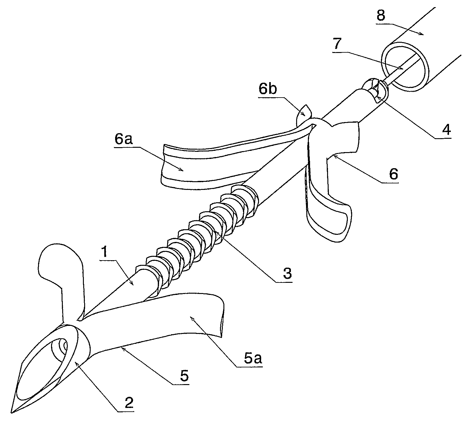
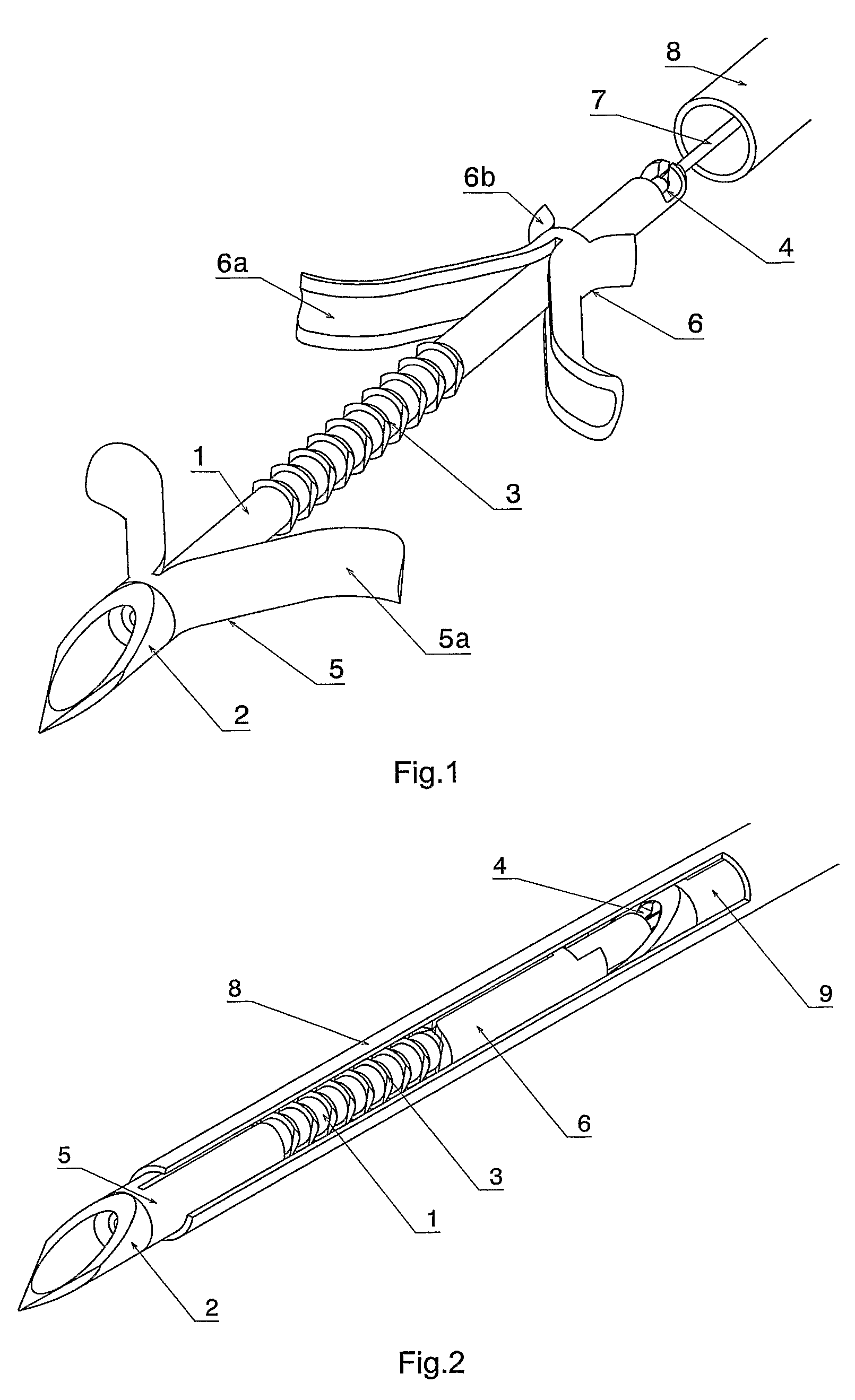
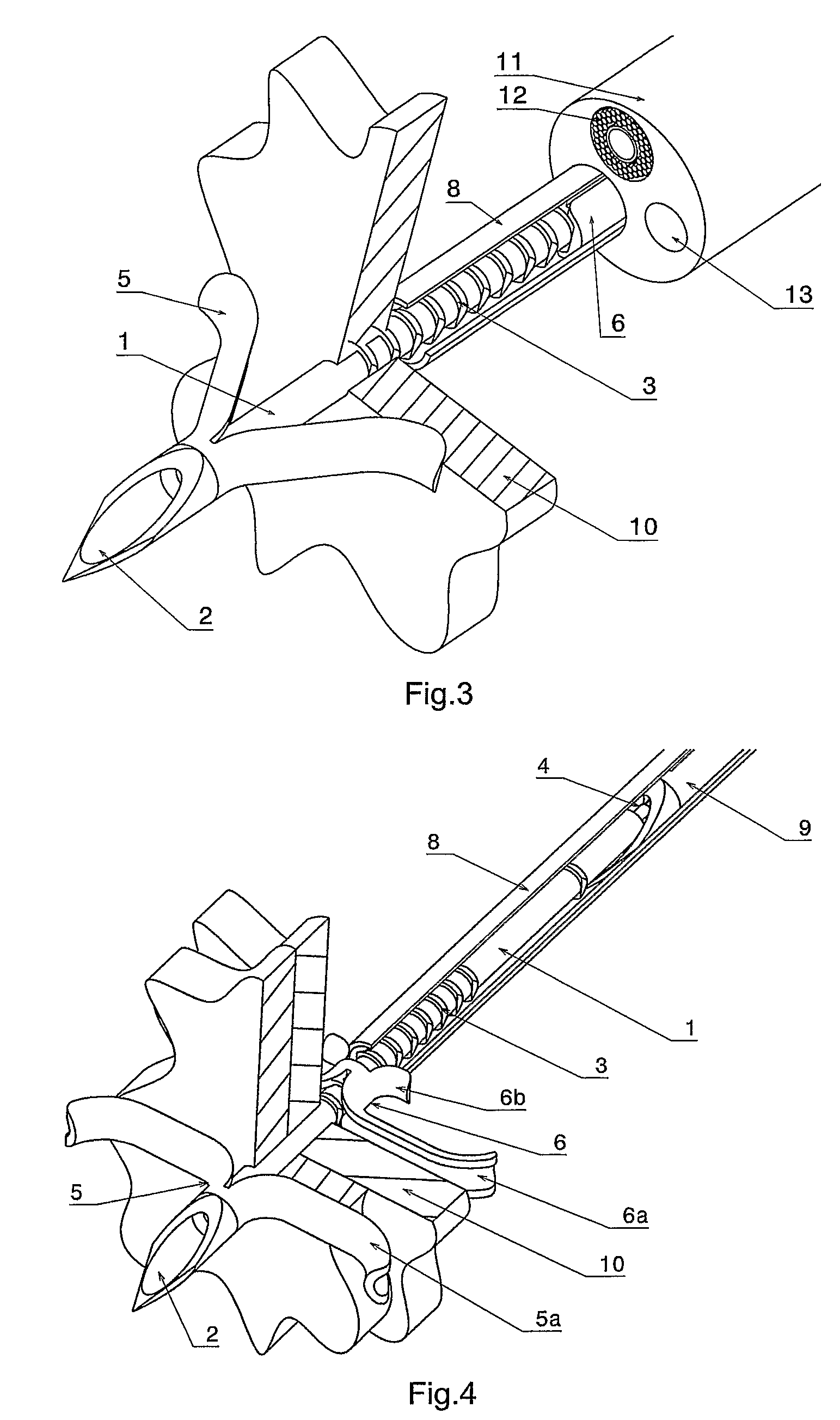
[0024]The rivet, as shown in its relaxed form in FIG. 1, consists essentially of three parts. A tubular carrier 1 is provided at the distal end with a tip 2, in the middle part with an arresting castellation 3 and, at the proximal end, with a coupling element 4. Two clips 5 and 6 are mounted on the carrier, the distal clip 5 being axially fixed and the proximal clip 6 being displaceable in the distal, axial direction and being held in the opposite direction by the arresting castellation. The distal clip consists of a basic, cylindrical body from which two arms 5a branch out. The proximal clip also consists of a basic, cylindrical body, from which two arms 6a, as well as two wings 6b spread out. Within the carrier, there is an actuating element 7 with a latch at the distal...


PUM


Abstract

A self-piercing blind rivet is provided for apposing biological tissue together with a device for setting the same, especially through the instrumental channel of a flexible endoscope. Without the help of further instruments, it is possible to fix, approximate and appose tissue layers, the contact force while connecting the tissue layers being adjustable. The rivets are stored in the setting device, so that they can be applied in order. The rivet and setting device enable the surgeon to combine tissue discontinuities conveniently by means of an endoscope. The surgical procedure is simplified further because the rivets can remain in the body.

Description

BACKGROUND OF THE INVENTION[0001]The invention relates to a self-piercing blind rivet for apposing biological tissue and to a device for setting the same, especially through the instrument channel of a flexible endoscope, in order to connect layers of tissue with an adjustable contacting force.[0002]Blind rivets are usually used to connect metal sheets, predominantly in aircraft construction. However, they can also be used for other materials, such as plastics, wood or leather. Their great advantage over other forms of rivets is that the parts to be joined have to be accessible only from one side, since there is no need for a bottom die to close the rivet. For most blind rivets, holes, which are one above the other for setting the rivet, must be drilled to start with. There are, however, also blind rivets, for example, in the EP 0 705 389 B1, which are self piercing in that they pierce the parts to be joined automatically during the setting process.[0003]In surgery also, there are f...

Claims


Application Information

Patent Timeline
IPC IPC(8): A61F2/08A61B17/04A61B17/068A61B17/08A61B17/68A61B17/064A61B19/00A61B17/00
CPCA61B2017/0408A61B2019/481A61B17/0643A61B2017/0647A61B2017/00004A61B2017/00867A61B17/0644A61B2017/0419A61B17/068A61B2090/08021
Inventor FEUSSNER, HUBERTUSHEINZL, JOACHIMHAUSMANN, ULRICHPASPA, ROBERT
Owner TECH UNIV MUNCHEN
Learn More