
A system for identifying focal liver lesions based on contrast-enhanced ultrasound

A contrast-enhanced ultrasound and focal technology, applied in the field of ultrasound, can solve problems such as tissue deformation, difference in feature recognition of contrast-enhanced images, and target disappearance in lesion areas

Inactive Publication Date: 2020-11-17
王瑛
View PDF7 Cites 0 Cited by
  • Summary
  • Abstract
  • Description
  • Claims
  • Application Information

AI Technical Summary

Problems solved by technology

In addition, the human visual system has inaccurate and uncertain defects in processing information, resulting in subjective differences in the recognition of contrast image features by different image analysts
In addition, when the number of recognition times is large, image analysts will inevitably experience visual fatigue, slow response, etc., and the error rate will increase.
At the same time, due to the data specifications, resolutions, and sizes collected by different devices, it is difficult to achieve uniformity, and when different doctors use the device probe to obtain images of contrast motion sequences, due to different techniques, probe vibration, human body movement and breathing. Causes tissue deformation and severe jitter, resulting in frequent shaking of the acquired sequence images, disappearance of targets in the lesion area, etc.
In addition, in the process of ultrasound imaging, due to the inhomogeneous characteristics of human cell vascular tissue, the superimposed scattered echoes will generate speckle noise, which will seriously affect the quality and resolution of contrast images, which is not conducive to the subsequent extraction and analysis of lesions Characteristics of objects in the zone

Method used


Image

  • A system for identifying focal liver lesions based on contrast-enhanced ultrasound
  • A system for identifying focal liver lesions based on contrast-enhanced ultrasound
  • A system for identifying focal liver lesions based on contrast-enhanced ultrasound

Examples


Embodiment

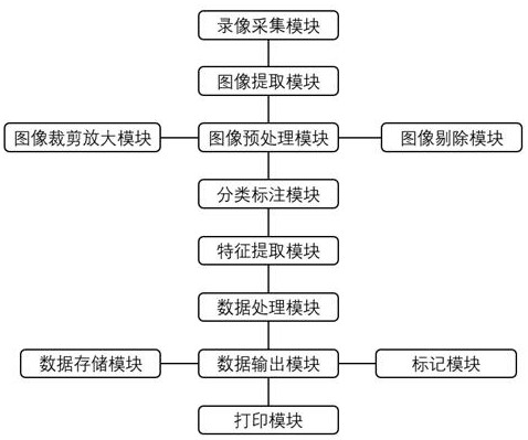
[0063] refer to figure 1 , a system for identifying focal liver lesions based on contrast-enhanced ultrasound, including a video acquisition module, an image extraction module, an image preprocessing module, a classification and labeling module, a feature extraction module, a data data module, and a data output module, wherein:

[0064] The video acquisition module is used to collect ultrasound contrast video from ultrasound contrast equipment;

[0065] The image extraction module is used to extract multiple frames of contrast-enhanced ultrasound images in time sequence from the contrast-enhanced ultrasound video;

[0066] The image preprocessing module is used to preprocess the contrast-enhanced ultrasound image data to obtain color-enhanced contrast data and divide the rectangular region of interest therein into several rectangular subregions of interest;

[0067] The classification and labeling module is used to classify and label several of the rectangular sub-regions of ...


PUM


Abstract

The invention discloses a system for identifying focal liver lesions based on contrast-enhanced ultrasonography. The system includes a video acquisition module, an image extraction module, an image pre-processing module, a classification labeling module, a feature extraction module, a data processing module, and a data output module, a mode based on combination of edge enhancement and color balance is used firstly for performing denoising and enhancement processing on a contrast image to enhance useful information of the image while retaining the color of the contrast image, then based on a gray level histogram of the image, feature extraction is performed on the contrast image to obtain reasonable quantitative parameters, so that the diagnostic errors caused by probe movement and lesion movement caused by breathing movements of a patient can be inhibited to the greatest extent, so that the features of the lesion is reflected scientifically, objectively, clearly, and effectively, so that accuracy rate and efficiency of feature identification of the focal liver lesions by a contrast-enhanced ultrasonography technology is improved, and thus a novel way is provided for the faster andbetter development of the contrast-enhanced ultrasonography technology.

Description

technical field [0001] The invention relates to the field of ultrasound technology, in particular to a system for identifying focal liver lesions based on contrast-enhanced ultrasound. Background technique [0002] Focal liver lesions are one of the common clinical liver lesions. Patients affected by the disease will continue to show symptoms such as liver function decline, and will cause serious damage to liver function after the disease progresses. Because most patients with focal liver lesions lack obvious lesions, it is difficult to find typical early symptoms, which makes misdiagnosis and missed diagnosis more likely to occur after seeing a doctor, which endangers the timing of effective treatment and even causes the disease to progress. endanger the health of the patient. Therefore, the most effective means of treating liver cancer is early detection and early treatment. Although CT, MRI, and especially PET imaging are effective in the early diagnosis of liver cancer...

Claims


Application Information

Patent Timeline
Patent Type & Authority Patents(China)
IPC IPC(8): A61B8/08
CPCA61B8/0833A61B8/481A61B8/5207A61B8/5215A61B8/5269A61B8/5276A61B8/5292
Inventor 王瑛赖铁强王宁张东海
Owner 王瑛
Learn More