Marking method, device and system of medical image

A medical image and marking method technology, applied in the medical field, can solve the problems of high work pressure, time-consuming and labor-intensive, low work efficiency, etc., and achieve the effect of improving efficiency and reducing the missed diagnosis rate.

Active Publication Date: 2020-02-14
INFERVISION MEDICAL TECH CO LTD
View PDF5 Cites 2 Cited by
  • Summary
  • Abstract
  • Description
  • Claims
  • Application Information

AI Technical Summary

Problems solved by technology

At present, most of the methods still use the mouse to slide layer by layer to view multiple two-dimensional images on the computer, and learn the position of the lesion area (region of interest) accordingly. Such work efficiency is obviously not high, and Due to the serious shortage of professional medical personnel, and the high requireme

Method used


Image

  • Marking method, device and system of medical image
  • Marking method, device and system of medical image
  • Marking method, device and system of medical image

Examples


Embodiment Construction

[0032] The following will clearly and completely describe the technical solutions in the embodiments of the present invention with reference to the accompanying drawings in the embodiments of the present invention. Obviously, the described embodiments are only some of the embodiments of the present invention, not all of them. Based on the embodiments of the present invention, all other embodiments obtained by persons of ordinary skill in the art without making creative efforts belong to the protection scope of the present invention.

[0033] Application overview

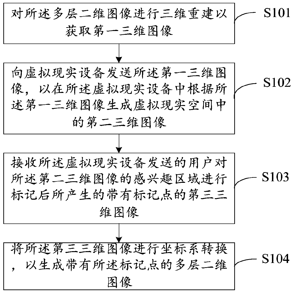
[0034]As mentioned above, the existing marking method of medical image inspection still uses the mouse to slide layer by layer to view multiple two-dimensional images on the computer by professional medical personnel, and obtains the position of the lesion area (region of interest) accordingly, but The work efficiency of detecting medical images through the existing marking method is obviously not high, and it also...


PUM


Abstract

The invention provides a marking method, device and system of a medical image. The marking method of the medical image comprises the following steps: performing three-dimensional reconstruction on a multi-layer two-dimensional image to acquire a first three-dimensional image; transmitting the first three-dimensional image to virtual reality equipment to generate a second three-dimensional image ina virtual reality space in the virtual reality equipment according to the first three-dimensional image; receiving a third three-dimensional image with a mark point, wherein the third three-dimensional image is generated after a user marks an interested area of the second three-dimensional image transmitted by the virtual reality equipment; and performing coordinate system conversion on the thirdthree-dimensional image to generate a multi-layer two-dimensional image with the mark point. The missed diagnosis rate of medical workers can be reduced and the medical image detection efficiency ofthe medical workers can be improved.

Description

technical field [0001] The invention relates to the field of medical technology, in particular to a marking method, device and system for medical images. Background technique [0002] Computed Tomography (CT for short) is a three-dimensional radiological medical image reconstructed after digital geometry processing. This technology mainly irradiates the human body through the rotation of X-rays on a single axis. Since different tissues have different absorption capabilities (or rejection rates) of X-rays, the three-dimensional technology of the computer can be used to reconstruct the tomographic image. By bit processing, the tomographic image of the corresponding tissue can be obtained, and the tomographic image can be stacked layer by layer to form a three-dimensional image. [0003] CT tomographic images are composed of multiple two-dimensional images stacked, which have three-dimensional characteristics. CT tomographic images are an important means and basis for judging ...

Claims


Application Information

Patent Timeline
IPC IPC(8): A61B6/03
CPCA61B6/032A61B6/466A61B6/5211
Inventor 余航陈宽王少康
Owner INFERVISION MEDICAL TECH CO LTD
Why Eureka
Try Eureka