
FCM-based diabetic retina image optic disk segmentation method

A technology of diabetic retina and optic disc, applied in the field of medical image processing

Pending Publication Date: 2020-03-17
HARBIN UNIV OF SCI & TECH
View PDF4 Cites 6 Cited by
  • Summary
  • Abstract
  • Description
  • Claims
  • Application Information

AI Technical Summary

Problems solved by technology

[0005] In view of the problems of the above research, the purpose of the present invention is to propose a FCM-based optic disc segmentation method for diabetic retinal images in order to reduce the time-consuming and accuracy problems caused by the large amount of calculation in the segmentation process.

Method used


Image

  • FCM-based diabetic retina image optic disk segmentation method
  • FCM-based diabetic retina image optic disk segmentation method
  • FCM-based diabetic retina image optic disk segmentation method

Examples


Embodiment Construction

[0045] In order to make the objectives, technical solutions and advantages of the present invention clearer, corresponding specific implementation plans are proposed, and the present invention is further described in detail.

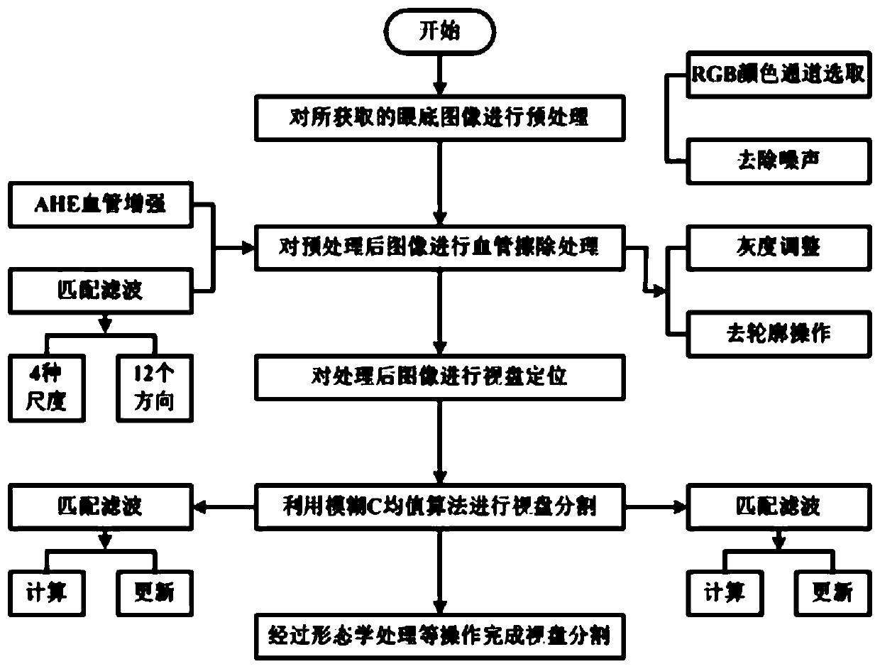
[0046] A method for segmenting the optic disc of a diabetic retinal image based on FCM, comprising the steps of:

[0047] S1: Obtain the original fundus image R'(x,y) to be processed, and perform image preprocessing on the image R'(x,y), which mainly includes the following parts: color channel analysis, noise removal, and preprocessed image R (x,y);

[0048] Further, the specific steps of S1 are:

[0049] S1.1: Select the G channel component image R”(x, y) of the original fundus image R’(x, y), which has a relatively high overall contrast in the RGB color channel, and a relatively high-definition optic disc brightness and blood vessel outline, as the processed image ;

[0050] S1.2: Use the mean filter method to reduce image noise, mainly to take the ...


PUM


Abstract

The invention discloses an FCM diabetic retina image optic disk segmentation method, and belongs to the technical field of image processing. The method mainly comprises the following steps: step 1, acquiring a to-be-processed original fundus image R '(x, y), and carrying out image preprocessing on the image R' (x, y); step 2, carrying out blood vessel processing, including blood vessel segmentation and blood vessel erasing, on the preprocessed image R (x, y), the blood vessel segmentation mainly comprising the following parts: blood vessel enhancement, matched filtering, gray scale adjustmentand contour elimination; step 3, carrying out optic disk positioning on the image R after the blood vessel is erased; and step 4, carrying out optic disk segmentation by utilizing an FCM (fuzzy C-means) algorithm. The problems of large calculation amount, long consumed time, low accuracy and the like are solved by using the FCM algorithm, and the overall efficiency of the optic disk segmentation process is improved.

Description

technical field [0001] The invention relates to the field of medical image processing, in particular to an FCM-based optic disc segmentation method for diabetic retinal images. Background technique [0002] Diabetes is a metabolic disease. In my country, the prevalence of diabetes is high, the treatment rate is low, and the incidence of complications caused by prolonged illness also increases. Among them, eye retinopathy is one of the most serious complications, which can cause human vision loss or even blindness. In the process of lesion detection on fundus images, the optic disc may interfere with the detection results, so the optic disc is selected for segmentation. [0003] The full name of the optic disc is the optic disc, also known as the optic nerve head, with a diameter of about 1.5mm. The optic nerve and blood vessels enter the eye from this area and extend to the periphery. In a normal color retinal fundus image, the optic disc generally appears as a nearly roun...

Claims


Application Information

Patent Timeline
Patent Type & Authority Applications(China)
IPC IPC(8): G06T7/00G06T7/11G06T7/90
CPCG06T7/0012G06T7/90G06T7/11G06T2207/30041G06T2207/30101G06T2207/20104
Inventor 高俊山韩婧邓立为
Owner HARBIN UNIV OF SCI & TECH
Learn More