CT image detection method and device, storage medium and electronic device

A detection method and CT image technology are applied in the field of image processing to achieve the effects of reducing workload, improving detection efficiency and avoiding missed detections

Inactive Publication Date: 2020-03-20
INFERVISION MEDICAL TECH CO LTD
View PDF10 Cites 5 Cited by
  • Summary
  • Abstract
  • Description
  • Claims
  • Application Information

AI Technical Summary

Problems solved by technology

[0004] In order to solve the above-mentioned technical problems, the present application proposes a detection method of CT images, by obtaining a single-layer two-dimensional image to be detected in a multi-layer two-dimensional image and continuous N layers of two-dimensional images on one side of the single-layer two-dimensional image to be detected 2D image and / or the continuous N-layer 2D image on the other side of the single-layer 2D image to be detected as the current image to be detected, where N is an integer greater than or equal to 1, and then the current image to be detected is input into the neural network model to generate The region of interest in the CT image to be detected can be replaced by manual detection of the region of interest in the CT image by using the neural network model, thereby greatly reducing the workload of medical staff in the radiology department, improving detection efficiency, and avoiding missed detection. In addition, considering that fractures and other positions are usually reflected in continuous multi-layer images, by detecting single-layer two-dimensional CT images and their upper and lower layers of continuous images, the detection results can be obtained by combining the upper and lower layers of images , to further improve the detection accuracy

Method used


Image

  • CT image detection method and device, storage medium and electronic device
  • CT image detection method and device, storage medium and electronic device
  • CT image detection method and device, storage medium and electronic device

Examples


Embodiment Construction

[0045] Hereinafter, exemplary embodiments according to the present application will be described in detail with reference to the accompanying drawings. Apparently, the described embodiments are only some of the embodiments of the present application, rather than all the embodiments of the present application. It should be understood that the present application is not limited by the exemplary embodiments described here.

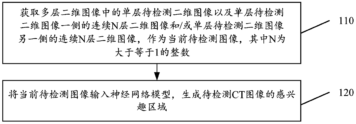
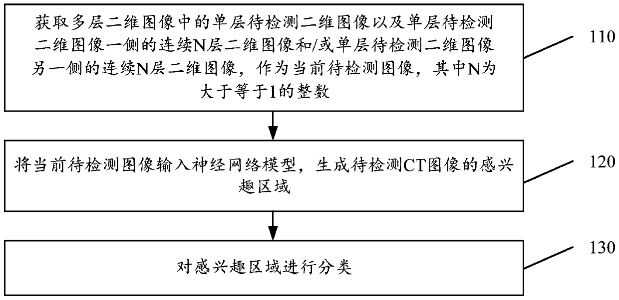
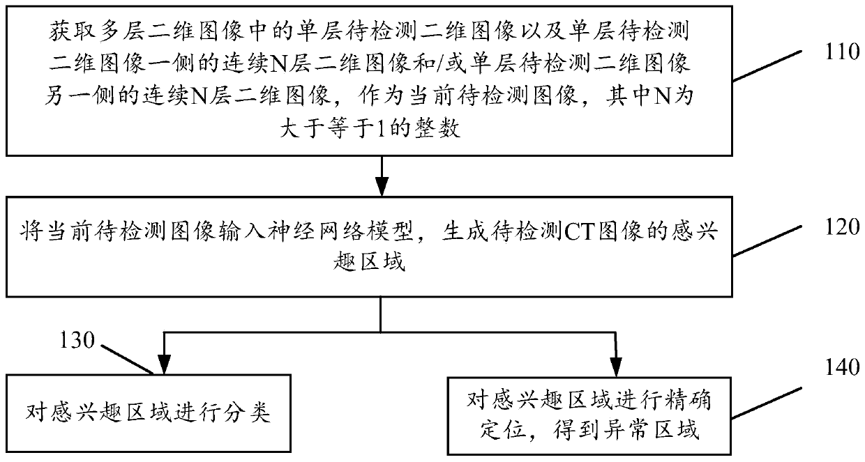
[0046] Application overview

[0047] CT tomographic images are composed of multiple two-dimensional images stacked, which have three-dimensional characteristics. CT tomographic images are an important means and basis for judging whether there is a fracture or other traumatic symptoms. At present, most of the methods still use professional medical personnel to manually view multiple two-dimensional images, and obtain the location of fractures and other lesions (regions of interest) accordingly. Such work efficiency is obviously not high, and due to a serious...


PUM


Abstract

The invention discloses a CT image detection method and device, a storage medium and an electronic device. The method comprises: acquiring a single-layer to-be-detected two-dimensional image and a single-layer to-be-detected two-dimensional image in multiple layers of two-dimensional images and continuous N layers of two-dimensional images on one side of the single-layer to-be-detected two-dimensional image and / or continuous N layers of two-dimensional images on the other side of the single-layer to-be-detected two-dimensional image as current to-be-detected images; wherein N is an integer greater than or equal to 1, and then generating a region of interest of the to-be-detected CT image by using the neural network model to replace manual detection of the region of interest in the CT image, thereby greatly reducing the workload of medical personnel, improving the detection efficiency, avoiding missing detection and improving the detection accuracy. Moreover, considering that positionssuch as fractures are usually reflected on continuous multi-layer images, a detection result is comprehensively obtained by detecting the multi-layer continuous images and combining upper-layer imagesand lower-layer images, so that the detection accuracy is further improved.

Description

technical field [0001] The present application relates to the field of image processing, and in particular to a CT image detection method, a detection device, a computer-readable storage medium, and electronic equipment. Background technique [0002] Computed Tomography (CT for short) is a three-dimensional radiological medical image reconstructed after digital geometry processing. This technology mainly irradiates the human body through the rotation of X-rays on a single axis. Since different tissues have different absorption capabilities (or rejection rates) of X-rays, the three-dimensional technology of the computer can be used to reconstruct the tomographic image. By bit processing, the tomographic image of the corresponding tissue can be obtained, and the tomographic image can be stacked layer by layer to form a three-dimensional image. [0003] Human CT tomography is an important means used to judge fractures in hospitals. The human body is fractured due to the actio...

Claims


Application Information

Patent Timeline
Patent Type & Authority Applications(China)
IPC IPC(8): G06T7/00
CPCG06T7/0012G06T2207/10081G06T2207/20104G06T2207/30008
Inventor 李玉才王少康陈宽
Owner INFERVISION MEDICAL TECH CO LTD
Why Eureka
Try Eureka