Rapid CT scanning method and system based on virtual stereotaxic image

A CT scanning and virtual stereo technology, applied in the field of medical imaging, can solve the problems of inaccurate positioning image area, excessive radiation exposure of patients, affecting scanning efficiency, etc., achieve accurate scanning area, save radiation dose and scanning time, and speed up scanning The effect of the process

Active Publication Date: 2020-04-03
NANJING ANKE MEDICAL TECH CO LTD
View PDF5 Cites 6 Cited by
  • Summary
  • Abstract
  • Description
  • Claims
  • Application Information

AI Technical Summary

Problems solved by technology

However, scanning the positioning image requires the rack to move from motion to stop until it is positioned at a specified angle before scanning. The acceleration, deceleration and positioning of the entire rack take time, so the preparation time for scanning is relatively long, which affects the scanning efficiency, especially Under the condition of applying dual positioning images, it cannot meet the needs of large-scale screening such as physical examination and screening
[0005] 3. In conventional CT scanning, due to the constraints of scanning efficiency and dose, often only one positioning image is used, and the scanning range is accurately determined according to one positioning image, which requires high clinical experience for doctors, and doctors who lack clinical experience The selected scout area may not be accurate enough, and the patient may be exposed to excessive radiation if scout scans are repeated

Method used


Image

  • Rapid CT scanning method and system based on virtual stereotaxic image
  • Rapid CT scanning method and system based on virtual stereotaxic image
  • Rapid CT scanning method and system based on virtual stereotaxic image

Examples


Embodiment Construction

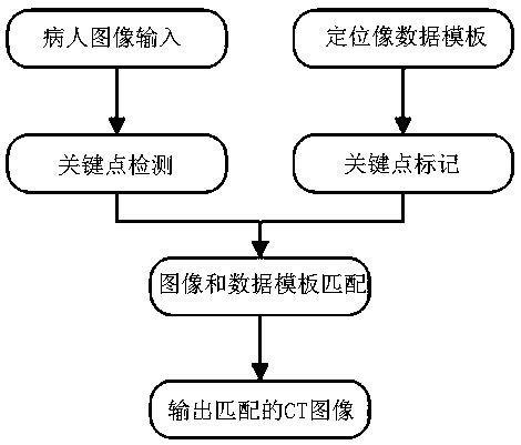
[0039] Such as Figure 1 to Figure 4 As shown, the present invention provides a fast CT scanning method and system based on virtual stereotaxic images, which uses rgb-d images to generate virtual stereotaxic images, specifically including steps 1) to 5).

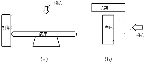
[0040] 1) Patient image input:

[0041] Such as figure 2 As shown, the input patient image is the patient image taken in two directions. Using optical image acquisition, a binocular vision system can be used to simultaneously acquire optical images and depth images (rgb-d images) in real time, saving time for mechanical movement.

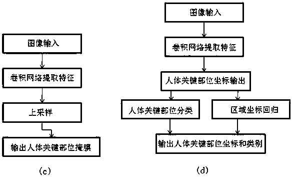
[0042] 2), key point detection:

[0043] The present invention can be used as image 3 The two network image keypoint detection methods shown work on patient images. As shown in Figure a, input an image, extract features through a convolutional network, and then output a mask of key parts of the human body through upsampling. As shown in Figure b, the image is input, features are extracted ...


PUM


Abstract

The invention discloses a rapid CT scanning method and system based on a virtual stereotaxic image. The rapid CT scanning method comprises the steps: inputting patient images which are photographed atdifferent angles and comprise a to-be-scanned region, and carrying out the key point detection of the patient images; inputting a positioning image data template and marking key points; performing matching calculation according to the key points in the patient image and the positioning image data template to obtain a three-dimensional virtual positioning image; and determining a CT scanning areaof the patient according to the three-dimensional virtual positioning image, and outputting a final CT image. The virtual positioning image generated by the optical image is used for replacing a traditional positioning image, and the time required for scanning the positioning image is shortened, and the radiation dose received by a patient is reduced, and the anatomical structure of the patient can be clearly displayed, and a doctor can more accurately position a scanning area, and the workload of the doctor is reduced.

Description

technical field [0001] The invention relates to the technical field of medical imaging, in particular to a fast CT scanning method and system based on a virtual stereotaxic image. Background technique [0002] Currently, in the CT scanning process, in order to select a scan area more accurately, it is necessary to scan a scout image first, and then select a scan area in the scout image. Such as Figure 5 As shown, the general CT process includes: patient registration; acquisition of horizontal and / or vertical positioning images according to scanning requirements; CT scanning. Doctors usually judge the scanning area of ​​the patient's positioning image based on experience, and the positioning image obtained by this method is not accurate. The Chinese patent "A Method and System for Intelligent Scanning Stereoscopic Monitoring" with the publication number CN110338835A discloses a method and system in which an auxiliary camera is used to capture an image of a patient to be sca...

Claims


Application Information

Patent Timeline
Patent Type & Authority Applications(China)
IPC IPC(8): G06T7/00G06K9/62
CPCG06T7/0012G06T2207/10081G06T2207/20081G06T2207/20084G06V10/751
Inventor 曾凯傅鹏何健
Owner NANJING ANKE MEDICAL TECH CO LTD
Why Eureka
Try Eureka