Three-dimensional hemoperfusion image generation method and device based on electric impedance imaging

A blood perfusion and electrical impedance measurement technology, which is applied in blood flow measurement, medical science, sensors, etc., can solve problems such as difficult to reflect blood perfusion, and achieve an effect that is conducive to image analysis and comparison

Active Publication Date: 2020-04-28
BEIJING HUARUI BOSHI MEDICAL IMAGING TECH CO LTD
View PDF7 Cites 9 Cited by
  • Summary
  • Abstract
  • Description
  • Claims
  • Application Information

AI Technical Summary

Problems solved by technology

Two-dimensional images are difficult to reflect the blood p

Method used


Image

  • Three-dimensional hemoperfusion image generation method and device based on electric impedance imaging
  • Three-dimensional hemoperfusion image generation method and device based on electric impedance imaging
  • Three-dimensional hemoperfusion image generation method and device based on electric impedance imaging

Examples


Embodiment Construction

[0028] The drawings are for illustration only and should not be construed as limiting the invention. The technical solutions of the present invention will be further described below in conjunction with the accompanying drawings and embodiments.

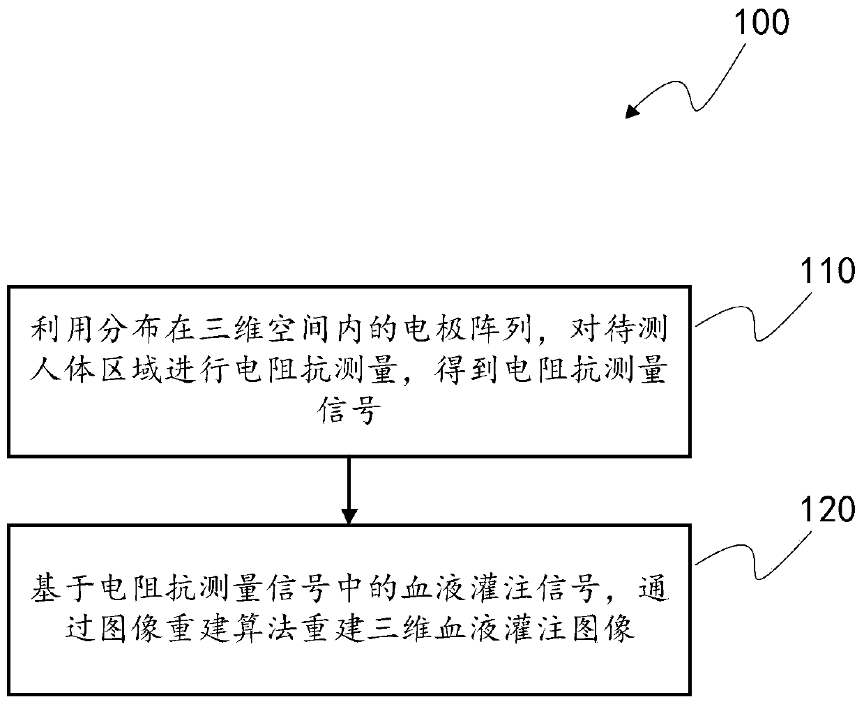
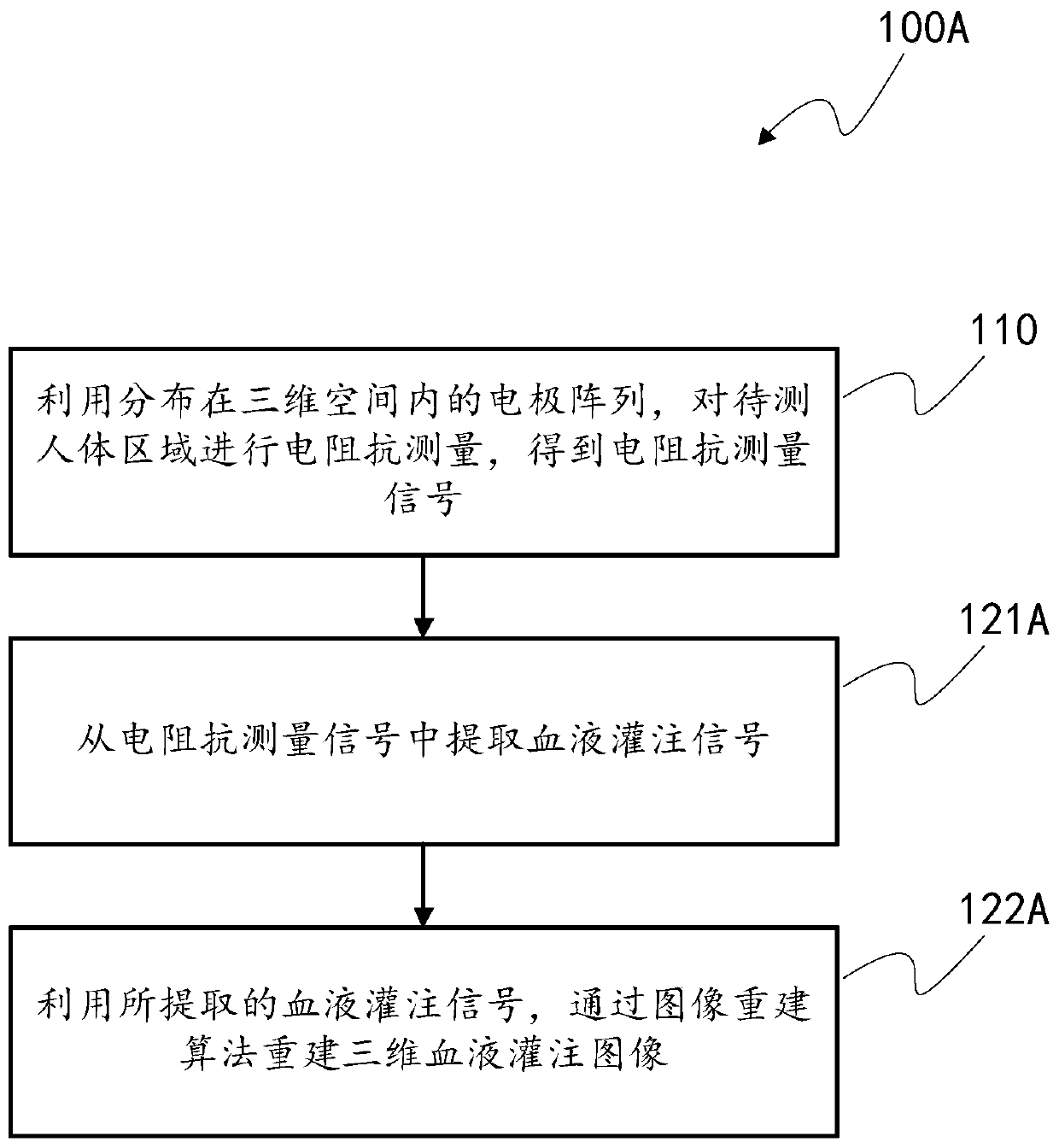
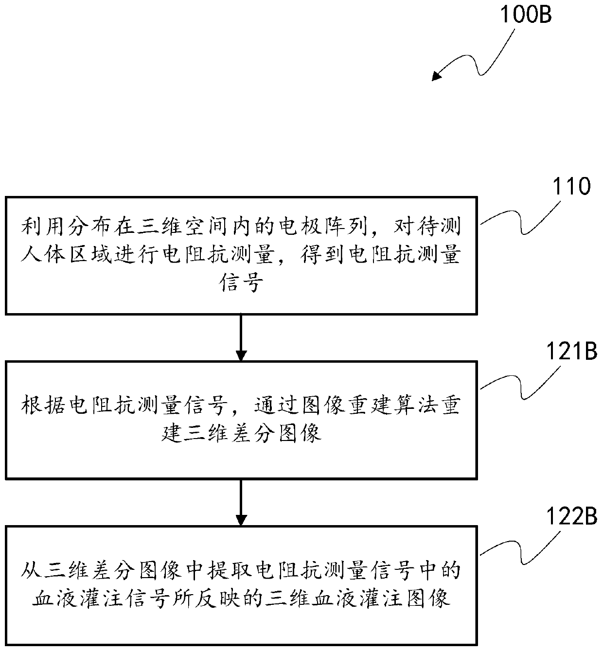
[0029] figure 1 is a schematic flowchart of a method 100 for generating a three-dimensional blood perfusion image based on electrical impedance imaging according to an embodiment of the present invention.

[0030] figure 1 The method 100 begins at step 110, where an electrical impedance signal of a human body is measured. Specifically, the electrode array distributed in the three-dimensional space is used to measure the electrical impedance of the area of ​​the human body to be measured to obtain electrical impedance measurement signals.

[0031] Electrical impedance measurements first require an array of electrodes to be fixed around the area of ​​the body to be measured. The electrode array includes several electrodes distribute...


PUM


Abstract

The invention provides a three-dimensional hemoperfusion image generation method and device based on electric impedance imaging. The method (100) comprises the following steps: performing electric impedance testing on human body areas to be tested by using electrode arrays distributed in a three-dimensional space so as to obtain electric impedance testing signals (110); and on the basis of hemoperfusion signals in the electric impedance testing signals, reconstructing a three-dimensional hemoperfusion image (120) by using an image reconstruction algorithm. Therefore, three-dimensional images of electric impedance variation caused by hemoperfusion can be generated, compared with two-dimensional images of the prior art, the three-dimensional images are capable of visibly reflecting hemoperfusion situations of one volume area in a three-dimensional space of a human body area, and image analysis and comparison and disease detection and diagnosis can be facilitated.

Description

technical field [0001] The present invention relates to electrical impedance imaging technology, and more particularly to a method and device for generating a three-dimensional blood perfusion image based on electrical impedance imaging. Background technique [0002] Electrical Impedance Tomography (EIT) is a non-invasive technique for reconstructing in vivo tissue images that targets the electrical resistivity distribution inside the human body or other organisms. The human body is a large bioelectrical conductor, and each tissue and organ has a certain impedance. When a local organ of the human body becomes diseased, the impedance of the local part must be different from that of other parts. Make a diagnosis. [0003] The electrical impedance imaging methods of the prior art can only reconstruct two-dimensional blood perfusion images. This two-dimensional image reflects the change of electrical impedance caused by blood perfusion in a section of the human body to be meas...

Claims


Application Information

Patent Timeline
IPC IPC(8): A61B5/053A61B5/026
CPCA61B5/0536A61B5/004A61B5/026A61B5/6802A61B5/725A61B2562/046
Inventor 张可张昕王谊冰于洋
Owner BEIJING HUARUI BOSHI MEDICAL IMAGING TECH CO LTD
Why Eureka
Try Eureka