Electrical impedance imaging apparatus and method

An electrical impedance imaging and equipment technology, applied in image generation, image data processing, 3D image processing, etc., can solve the inconvenience of disease detection and diagnosis, cannot display lung ventilation images and perfusion images at the same time, and cannot reflect biological tissue. Or the three-dimensional structure information of organs, etc., to achieve the effect of being conducive to image analysis and comparison, improving speed, and improving sensitivity

Pending Publication Date: 2019-06-11
BEIJING HUARUI BOSHI MEDICAL IMAGING TECH CO LTD
View PDF0 Cites 11 Cited by
  • Summary
  • Abstract
  • Description
  • Claims
  • Application Information

AI Technical Summary

Problems solved by technology

However, two-dimensional images cannot reflect the three-dimensional structural information of biological tissues or organs, which will cause inconvenience to the detection and diagnosis of diseases
[0007] In addition, current electrical impedance imaging devices either can only display images of ventilation of the lungs, or one of the images of ventilation and perfusion of the lungs alone, and cannot display both ventilation and perfusion images of the lungs at the same time

Method used


Image

  • Electrical impedance imaging apparatus and method
  • Electrical impedance imaging apparatus and method
  • Electrical impedance imaging apparatus and method

Examples


Embodiment Construction

[0025] The drawings are for illustration only and should not be construed as limiting the invention. The technical solutions of the present invention will be further described below in conjunction with the accompanying drawings and embodiments.

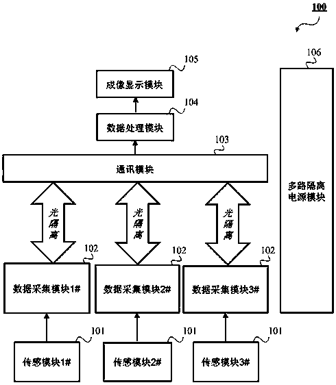
[0026] figure 1 is a compositional block diagram of the electrical impedance imaging device 100 according to the embodiment of the present invention.

[0027] Such as figure 1 As shown, according to the embodiment of the present invention, the electrical impedance imaging device 100 is generally composed of a sensing module 101 , a data acquisition module 102 , a communication module 103 , a data processing module 104 , an imaging display module 105 and a power supply module 106 . Wherein, the sensor module 101 and the data acquisition module 102 are electrically isolated from the communication module 103 , the data processing module 104 , the imaging display module 105 and the power supply module 106 .

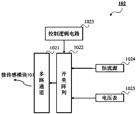
[0028] The sensing module ...


PUM


Abstract

An electrical impedance imaging apparatus and method are provided. The electrical impedance imaging device (100) is generally composed of a sensing module (101), a data acquisition module (102), a communication module (103), a data processing module (104), an imaging display module (105) and a power supply module (106). The electrical impedance imaging device (100) of the invention is applied to medical imaging, an in-vivo electrode can be used for carrying out simultaneous multi-frequency excitation and measurement on a to-be-measured organism tissue; three-dimensional image reconstruction iscarried out by using a measured complex voltage signal, and ventilation and perfusion images can be displayed in real time at the same time, so that the number of acquired data is increased, the dataacquisition speed is increased, the sensitivity of the measurement signal to the conductivity of in-vivo tissues is improved, and image analysis comparison and disease detection and diagnosis are facilitated.

Description

technical field [0001] The invention relates to electrical impedance imaging technology, and more particularly to a three-dimensional simultaneous multi-frequency complex electrical impedance imaging device and method applied to medical imaging. Background technique [0002] Electrical impedance imaging (Electrical Impedance Tomography, EIT) is a non-invasive technique that aims at the electrical resistivity distribution inside the human body or other organisms to reconstruct tissue images in the body. The human body is a large bioelectrical conductor, and each tissue and organ has a certain impedance. When a local organ of the human body becomes diseased, the impedance of the local part must be different from that of other parts. Make a diagnosis. [0003] Current electrical impedance imaging equipment uses external electrodes for data collection, that is to say, the electrodes are all arranged on the outer surface of the body to be measured. The signals thus acquired are...

Claims


Application Information

Patent Timeline
Patent Type & Authority Applications(China)
IPC IPC(8): A61B5/00A61B5/053
CPCA61B5/0536A61B5/6846A61B5/743A61B2560/0223A61B2562/046G06T15/00G06T2210/41
Inventor 张昕王谊冰张可于洋
Owner BEIJING HUARUI BOSHI MEDICAL IMAGING TECH CO LTD
Why Eureka
Try Eureka