
Robust interactive medical image segmentation method

A medical image, interactive technology, applied in the field of robust interactive medical image segmentation, can solve the problems of low contrast, poor effect, uneven intensity distribution, etc., achieve strong recognition ability, overcome the effect of low contrast

Pending Publication Date: 2020-05-15
SHANDONG UNIV
View PDF5 Cites 1 Cited by
  • Summary
  • Abstract
  • Description
  • Claims
  • Application Information

AI Technical Summary

Problems solved by technology

[0003] For retinal OCT images, the intensity distribution is extremely uneven, usually manifested as low contrast, and severe speckle noise, so the effect of global segmentation of lesions is not good, and it is difficult to segment and identify small lesions
The traditional region-based active contour model relies on the global information of the image. When dealing with images with uneven grayscale intensity, the same category cannot be effectively identified based on global information when the grayscale is different due to uneven grayscale.

Method used


Image

  • Robust interactive medical image segmentation method
  • Robust interactive medical image segmentation method
  • Robust interactive medical image segmentation method

Examples


Embodiment 1

[0043] A robust interactive medical image segmentation method, comprising the following steps:

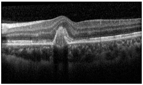
[0044](1) The original OCT image without processing, such as figure 1 As shown, the original OCT image is first regularized, and the purpose of the regularization is to normalize the pixel values ​​of the original image to the range of [0,1]; then the fractional differential enhancement is performed on the image, the purpose is to reduce Influenced by speckle noise, the specific process of fractional differential enhancement is as follows:

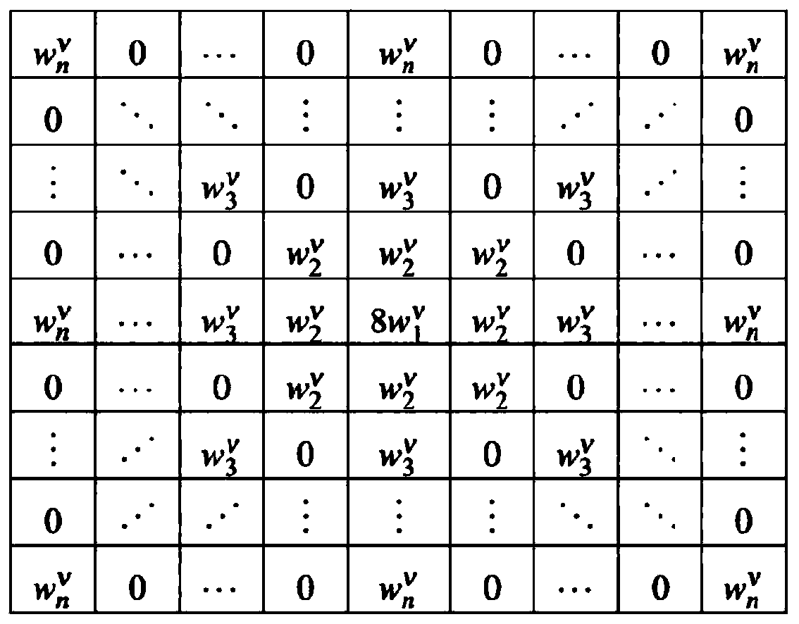
[0045] First, the n×n fractional differential enhancement mask is obtained using a discrete method, such as figure 2 As shown, any element Γ(-v+1) represents the gamma function of (-v+1), Γ(-v+i+1) represents the gamma function of (-v+i+1), i! Represents the factorial of i, where n=5, v=0.2;

[0046] Then, the image is convolved with an n×n fractional differential enhancement mask to complete the fractional differential enhancement of the...


PUM


Abstract

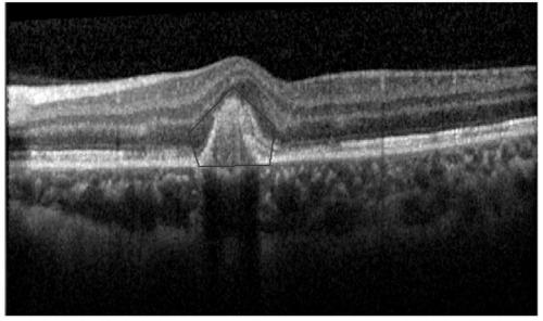
The invention relates to a robust interactive medical image segmentation method. The segmentation method comprises the following steps: (1) pre-segmenting a window where a retinal lesion is located inan OCT image; (2) setting two fixed foot points in the window to form a to-be-segmented window; and performing iterative contraction by using the active contour model in the to-be-segmented window tocomplete segmentation. According to the image segmentation method based on the active contour model, the segmentation range of a retina OCT image with high speckle noise is reduced through manual windowing so as to eliminate the influence of speckle noise in a background area on the model and accelerate the focus segmentation process. Inaccurate segmentation caused by the lightproof characteristic of the focus can be overcome through the foot points; in combination with fractional differential enhancement and a local fuzzy energy functional, an active contour model is proposed, so focus segmentation can be completed robustly and efficiently.

Description

technical field [0001] The invention relates to a robust interactive medical image segmentation method, which belongs to the technical field of image processing. Background technique [0002] Retinal optical coherence tomography (OCT) images are an effective basis for the diagnosis of many ophthalmic diseases and other diseases, such as diabetic retinopathy, macular edema, and calcification generally appear as corresponding plaques in OCT images. Each layer of the retinal image has its own specific structural features, varying in size, shape and distribution. The signal and noise models of each layer are different. Pathological structures with distinct features generally appear at specific levels. For example, the tympanic membrane structure appears just below or just above the retinal pigment epithelium (RPE). Hyperreflective foci associated with age-related macular degeneration are not expected to occur in the nerve fiber layer (NFL). In clinical diagnosis, relevant le...

Claims


Application Information

Patent Timeline
IPC IPC(8): G06T7/00G06T7/10
CPCG06T7/0012G06T7/10G06T2207/10101G06T2207/30041Y02T10/40
Inventor 付树军谢时灵赵志刚李玉亮唐荣王凤苓
Owner SHANDONG UNIV
Learn More