A vein imaging device and a method for generating a three-dimensional panoramic model thereof

A camera device and vein technology, applied in 3D modeling, image analysis, image enhancement, etc., can solve the problems of complex calculation process, increased implementation difficulty, and inability to reconstruct vein images in 3D, so as to realize 3D reconstruction, improve imaging quality, Simplifies calculating complex effects

Active Publication Date: 2022-04-12
XIEHE HOSPITAL ATTACHED TO TONGJI MEDICAL COLLEGE HUAZHONG SCI & TECH UNIV
View PDF6 Cites 0 Cited by
  • Summary
  • Abstract
  • Description
  • Claims
  • Application Information

AI Technical Summary

Problems solved by technology

[0003] The invention application with the application number 201610980034.0 discloses a device and method for three-dimensional reconstruction of human superficial veins based on binocular stereo vision. The near-infrared light source and the camera are respectively placed on both sides of the acquisition object, effectively avoiding the skin surface structure and texture. The reflection of the light source can improve the imaging quality, but the fusion process of near-infrared light source and camera imaging is not easy to calibrate, and the calculation process is complicated, which increases the difficulty of implementation
[0004] The invention application with the application number of 201811642253.3 discloses a method and device for obtaining finger vein image enhancement. A low-cost camera can be used to obtain finger vein images irradiated with near-infrared rays. After obtaining the finger vein images, pass through a two-dimensional symmetrical mask matrix The low-frequency matrix coefficients of discrete wavelet transform pre-process the finger vein image, and obtain the texture feature of the finger vein by detecting the valley points of the processed finger vein image, which can identify the position of the vein image, but cannot perform three-dimensional reconstruction of the vein image. Get the skin depth where the vein is

Method used


Image

  • A vein imaging device and a method for generating a three-dimensional panoramic model thereof
  • A vein imaging device and a method for generating a three-dimensional panoramic model thereof
  • A vein imaging device and a method for generating a three-dimensional panoramic model thereof

Examples


Embodiment Construction

[0061] The present invention will be further described in detail below in conjunction with the accompanying drawings, so that those skilled in the art can implement it with reference to the description.

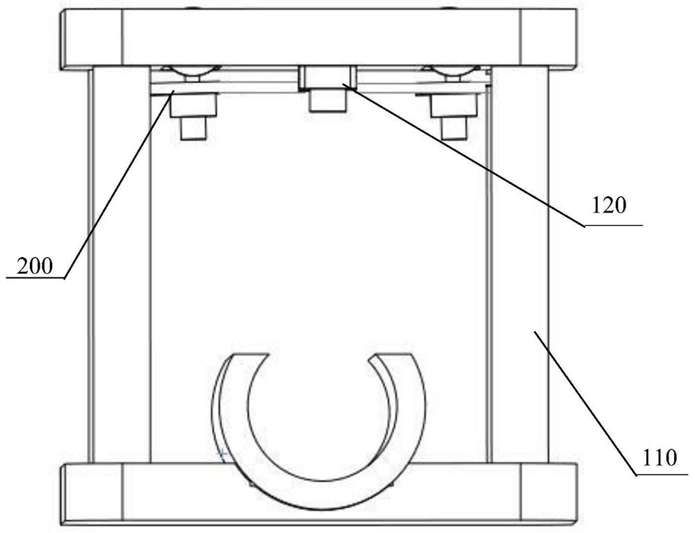
[0062] Such as figure 1 As shown, the vein developing camera device provided by the present invention includes: a frame 110 , a camera 120 and a binocular infrared camera device 200 .

[0063] As a preference, the frame 110 is a frame structure, the camera 120 is arranged on the top of the frame 110, and can take visible light two-dimensional images of the patient's area to be developed, and the bottom of the frame 110 has a calibration structure, which can calibrate the area of ​​the patient to be developed. The position is such that the area of ​​the patient to be developed is set coaxially with the camera 120 .

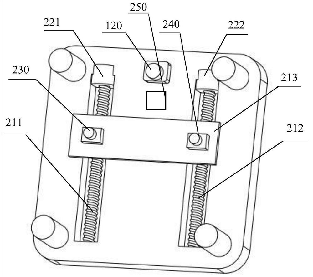
[0064] Such as figure 2 As shown, the first lead screw 211 is rotatably supported on the top of the frame 110; the second lead screw 212 is rotatably supported...


PUM


Abstract

The present invention provides a photographic device for developing veins, comprising: a frame; a camera, which is arranged on the top of the frame, capable of photographing two-dimensional visible light images of the patient's area to be developed; a synchronous driving device, which is respectively connected to the first lead screw and the second lead screw, which can drive the first lead screw and the second lead screw to rotate synchronously; the first infrared camera, which is arranged at one end of the synchronous frame, can photograph the area of ​​the patient to be developed to obtain the first two-dimensional Infrared image; the second infrared camera, which is arranged at the other end of the synchronous frame, can photograph the area of ​​the patient to be developed to obtain a second two-dimensional infrared image; the first laser, which is arranged on the first infrared camera, can The first visible light spot is formed on the patient's surface; the second laser, which is set on the second infrared camera, can form the second visible light spot on the patient's surface; the image re-fusion processing module also provides a generation of a 3D panoramic model of vein development photography method.

Description

technical field [0001] The invention relates to the technical field of vein development recognition, in particular to a vein development camera device and a method for generating a vein development camera three-dimensional panoramic model. Background technique [0002] Infrared imaging of veins plays an important role in medical assistance and biometric identification. In terms of medical treatment, venous imaging technology has been developed rapidly. According to relevant data, the success rate of normal adult venipuncture for the first time is 73%; the failure rate of the first two venipuncture for children is 58%; Generally speaking, the probability of "missing the needle" during venipuncture is 23-28%. Since there are many superficial subcutaneous tissues in the human body, it will be affected during the venipuncture process, such as: blood vessel depth, fat layer thickness, skin pigment content, etc. Affected by many factors, inexperienced or even experienced medical ...

Claims


Application Information

Patent Timeline
Patent Type & Authority Patents(China)
IPC IPC(8): G06T17/00G06T7/136
CPCG06T17/00G06T7/136G06T2207/10012G06T2207/10048
Inventor 叶霖韩斌陈学东杨新龙绍军
Owner XIEHE HOSPITAL ATTACHED TO TONGJI MEDICAL COLLEGE HUAZHONG SCI & TECH UNIV
Why Eureka
Try Eureka