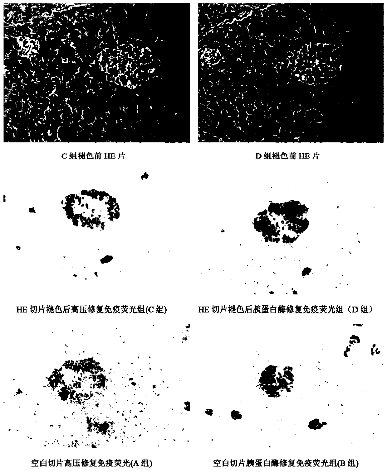
Method for immunofluorescence staining after HE slice fading

An immunofluorescence staining and sectioning technology, applied in the field of medical detection, can solve the problems of unfavorable original HE sectioning, comparative research, small tissue pieces and inability to tissue section immunofluorescence staining, etc.

Inactive Publication Date: 2020-06-19
GUANGDONG LAB ANIMALS MONITORING INST
View PDF2 Cites 1 Cited by
  • Summary
  • Abstract
  • Description
  • Claims
  • Application Information

AI Technical Summary

Problems solved by technology

Sometimes the diagnosis or further research of lesions under HE staining requires the assistance of immunofluorescence staining. At this time, re-cutting of the paraffin block is often chosen, but the re-cut section is not the same tissue slice as that observed by HE, and the position to be observed has already been or more. Some changes have occurred, which is not conducive to accurate comparison research with the original HE slices. What's more, some tissue blocks are small and cannot be cut out for immunofluorescence staining after heavy sectioning.

Method used


Image

  • Method for immunofluorescence staining after HE slice fading

Examples


Embodiment

[0032] Experimental animals and experimental environment

[0033] [Ordinary level] 3 crab monkeys with food, provided by Guangdong Chunsheng Biotechnology Development Co., Ltd. and raised in the general environment animal facility of Guangdong Chunsheng Biotechnology Development Co., Ltd. Use ethics committee approval. The animal breeding sites carry out daily care and management of animals in strict accordance with the regulations and requirements of the International Laboratory Animal Evaluation and Certification Management Committee.

[0034] Instruments and reagents

[0035] The instruments used include Leica TP1020 automatic dehydrator, Leica EG1150H+C paraffin embedding machine, Leica RM2255 automatic rotary microtome, Leica ST5020+SV5030 automatic staining and sealing machine, Leica DM3000 upright fluorescence microscope.

[0036] The reagents used include discoloration agent 1% hydrochloric acid ethanol differentiation solution, insulin antigen (Gene Tex company), PB...


PUM


Abstract

The invention discloses a method for immunofluorescence staining after HE slice fading. A slice after HE staining is provided. The method comprises the following steps: soaking a slice subjected to HEstaining by using xylene, and removing cover glass and neutral gum; carrying out hydrating and fading with gradient alcohol, and soaking the processed slice in a 1% of hydrochloric acid ethanol differentiation solution prepared from 75% alcohol for 1 min; after drying in air, putting the processed slice into a 1% of hydrochloric acid alcohol differentiation solution for 1 minute, and repeating the third step for multiple times until the color of the slice is completely faded; slowly washing the processed slice with tap water and then carrying out high-pressure repairing; and carrying out immunofluorescence staining on the slice after high-pressure repair. The method disclosed by the invention has the beneficial effects that the comparison under the same antigen repair method shows that the antigen expression intensity of the dyed slice after HE fading is equivalent to that of the slice directly dyed by a blank sheet, which indicates that the INS antigen is better preserved by the method disclosed by the invention.

Description

technical field [0001] The invention relates to the technical field of medical detection, in particular to a method for performing immunofluorescent staining on HE slices after fading. Background technique [0002] Immunofluorescence detection technology has the advantages of strong specificity, high sensitivity, and good practicability [1], and is an important auxiliary technology in pathological detection. HE staining is a routine staining method for pathological detection, and some meaningful morphological changes were found in HE staining. Sometimes the diagnosis or further research of lesions under HE staining requires the assistance of immunofluorescence staining. At this time, re-cutting of the paraffin block is often chosen, but the re-cut section is not the same tissue slice as that observed by HE, and the position to be observed has already been or more. Some changes have occurred, which is not conducive to accurate comparison with the original HE slices. What's m...

Claims


Application Information

Patent Timeline
Patent Type & Authority Applications(China)
IPC IPC(8): G01N1/30G01N33/533
CPCG01N1/30G01N33/533
Inventor 高洪彬陈梅丽王希龙梁十郭扬清吴玉娥贾欢欢龚宝勇刘婵刘晓霖麦冬梅
Owner GUANGDONG LAB ANIMALS MONITORING INST
Why Eureka
Try Eureka