Tomography and Image-Guided Radiation Therapy Devices

A radiation therapy and image guidance technology, applied in the field of medical imaging and radiation therapy guidance, can solve the problems of low spatial resolution, low detection sensitivity, and large scattering artifacts of CT imaging, avoid X-rays entering, improve detection sensitivity, The effect of improving accuracy

Active Publication Date: 2021-03-05
TSINGHUA UNIV
View PDF7 Cites 0 Cited by
  • Summary
  • Abstract
  • Description
  • Claims
  • Application Information

AI Technical Summary

Problems solved by technology

[0008] At present, the CT imaging and image-guided radiotherapy devices based on PET detectors have two main limiting factors for the CT imaging quality: 1) The single pixel unit of PET detector is generally larger than the single pixel unit of CT detector; 2) PET detector Detectors are generally not descatter collimated
These limiting factors will result in low spatial resolution of CT imaging, large scattering artifacts, and low detection sensitivity

Method used


Image

  • Tomography and Image-Guided Radiation Therapy Devices
  • Tomography and Image-Guided Radiation Therapy Devices
  • Tomography and Image-Guided Radiation Therapy Devices

Examples


Embodiment Construction

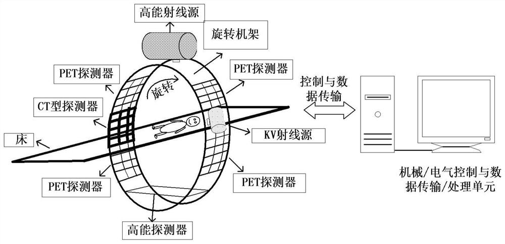
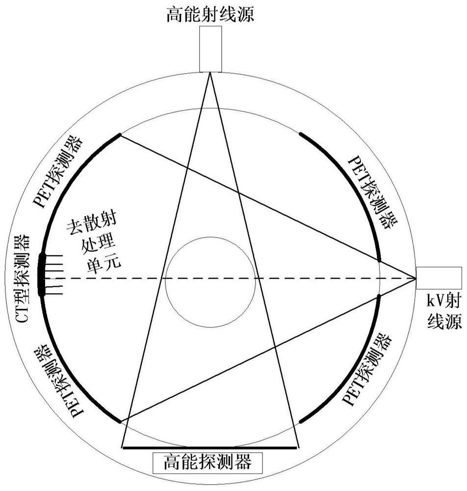
[0051] In the prior art, tomographic imaging and image-guided radiotherapy devices based on PET detectors have the defects of low spatial resolution of CT imaging, large scattering artifacts, and low detection sensitivity. In view of this,

[0052] In order to make the object, technical solution and advantages of the present invention clearer, the present invention will be described in further detail below in conjunction with specific embodiments and with reference to the accompanying drawings.

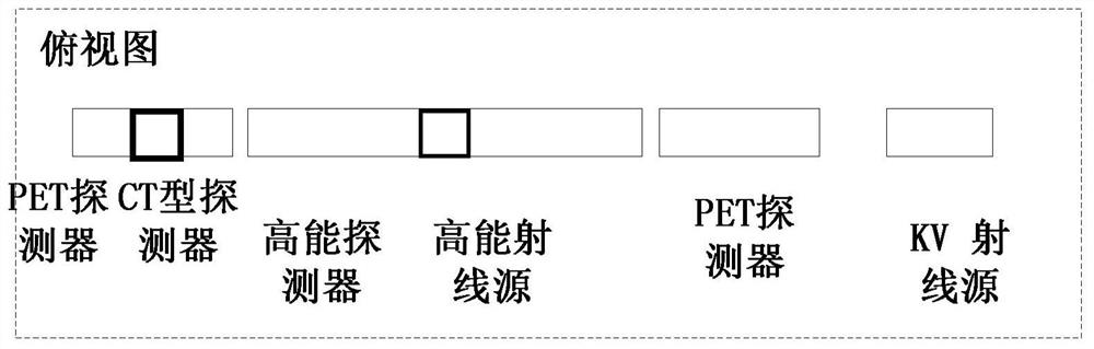
[0053] An embodiment of the present invention provides a tomographic imaging and image-guided radiotherapy device, including: at least one high-energy ray source for radiotherapy; a first detector and a second detector placed oppositely, and the first detector is located close to KV On one side of the ray source, the second detector is located on the side away from the KV ray source, the second detector includes at least two sections of PET detectors and a CT-type detector located betw...


PUM


Abstract

A device for tomographic imaging and image-guided radiotherapy, including a high-energy ray source; a first detector and a second detector, the second detector includes a PET detector and a CT-type detector located between the PET detectors; the first At least one kilovolt KV ray source for medical diagnosis is placed on the detector, the inside of the first detector or the outside of the first detector to generate KV ray; the second detector receives KV ray and performs KVCT imaging; the first detection The detector and the second detector are also used to receive gamma rays emitted by the object for PET imaging; the high-energy ray source generates high-energy rays for radiotherapy of the object; KVCT imaging and / or PET imaging are used for assisting and / or To guide radiation therapy to an object. The invention adopts CT detector and PET detector at the same time, improves the detection sensitivity of the device, improves the spatial resolution of KVCT imaging, and reduces scattering artifacts.

Description

technical field [0001] The invention relates to the fields of medical imaging and radiotherapy guidance, in particular to a tomographic imaging and image-guided radiotherapy device. Background technique [0002] Radiation therapy (Radiation Therapy) is currently one of the main ways to treat malignant tumors. In radiation therapy, image guidance (Image Guidance) is one of the key means to ensure high-precision treatment. Radiation therapy guidance is essentially the use of medical imaging to obtain accurate body position information of the patient before or during treatment, reducing the treatment errors caused by the patient's body placement, heartbeat, breathing and other organ movements, thereby improving or ensuring radiation therapy. the accuracy. Currently, the most common radiation therapy is the use of high-energy X-rays at the megavolt (MV) level, and the most common radiation therapy guidance is computed tomography (CT) based on kilovolt (KV) X-rays. In recent y...

Claims


Application Information

Patent Timeline
Patent Type & Authority Patents(China)
IPC IPC(8): A61B6/03A61N5/10
CPCA61B6/037A61B6/5205A61B6/5211A61B6/5258A61N5/1039A61N5/1049A61N2005/1052
Inventor 高河伟陈志强张丽邢宇翔
Owner TSINGHUA UNIV
Why Eureka
Try Eureka