Auxiliary support for fluorescein fundus angiography examination

A technology of auxiliary bracket and fluorescein, which is applied in the direction of ophthalmoscope, etc., can solve the problems of inconvenient inspection personnel, lack of auxiliary bracket, injection of contrast agent, etc., and achieve the effect of convenient contrast agent injection, reducing space occupation and improving comfort

Pending Publication Date: 2020-07-14
柳州市柳铁中心医院
View PDF0 Cites 0 Cited by
  • Summary
  • Abstract
  • Description
  • Claims
  • Application Information

AI Technical Summary

Problems solved by technology

[0003] In view of the above, it is necessary to provide an auxiliary support for fundus fluorescein angiography examination, which is used to solve the problems caused by the small desktop of the existing f...

Method used


Image

  • Auxiliary support for fluorescein fundus angiography examination
  • Auxiliary support for fluorescein fundus angiography examination
  • Auxiliary support for fluorescein fundus angiography examination

Examples


Embodiment

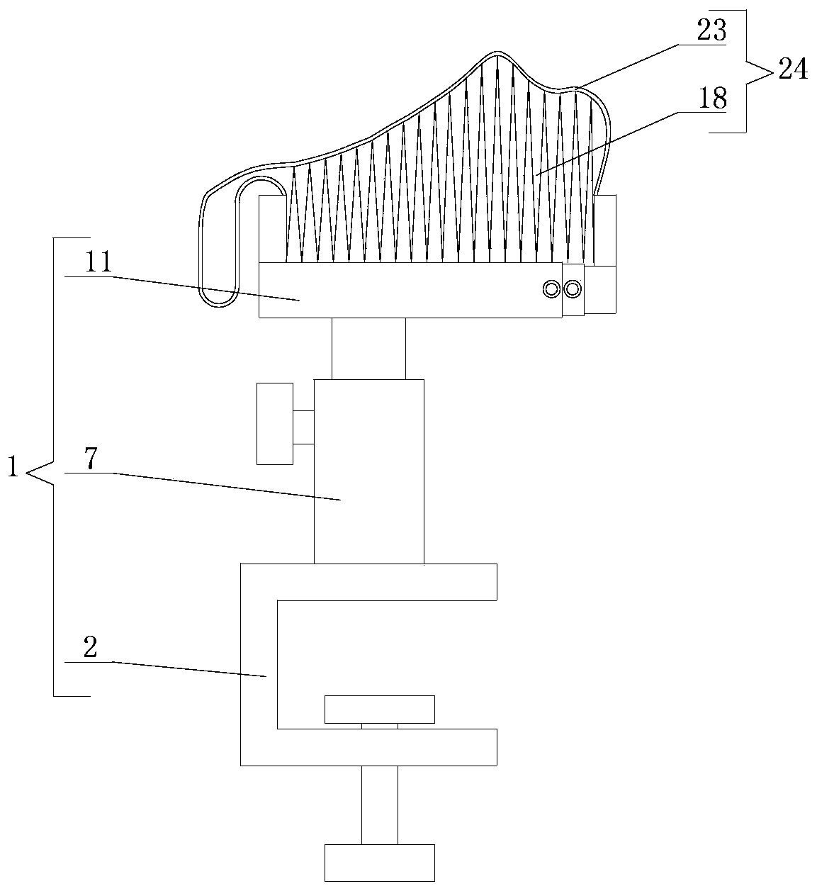
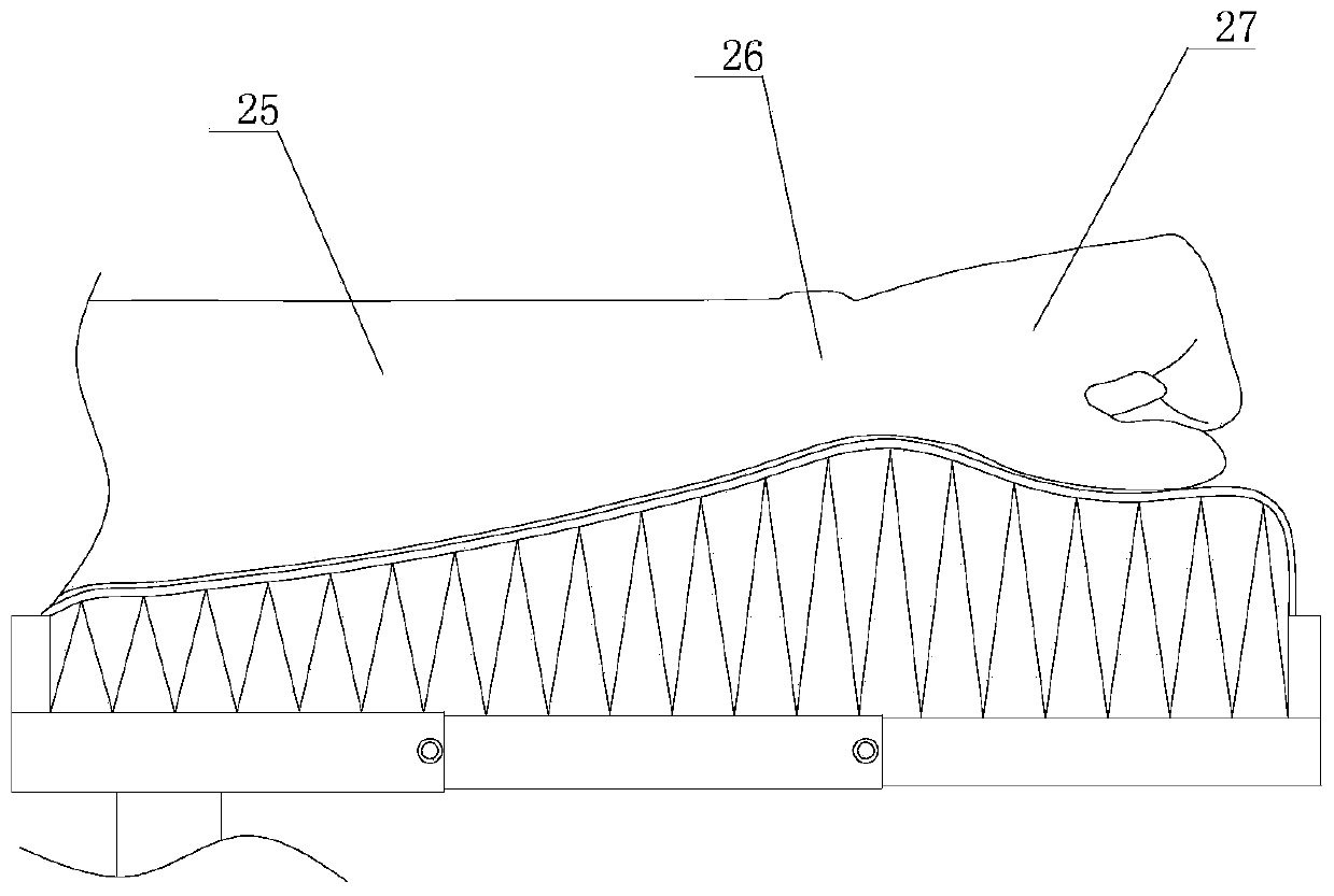
[0032] see Figure 1-4 , an auxiliary frame for fluorescein angiography examination, including a support frame 1 and a foldable forearm pad 24 .

[0033] The support frame 1 includes a fixing clip 2 , a support rod 7 and a telescopic plate 11 . The fixing clip 2 includes a concave clip body 3 , a first thumb screw 5 and a splint 6 . The notch 4 of the concave clamp body 3 is arranged horizontally, and the first thumb screw 5 vertically passes through the bottom of the concave clamp body 3 and is threadedly connected with the concave clamp body 3 . The splint 6 is located in the concave clamp body 3 and is fixedly connected to the upper end of the first thumb screw 5 . Preferably, the upper end of the first thumbscrew 5 is rotationally connected with the splint 6 , so that the splint 6 will not rotate with the first thumbscrew 5 , but will only move up and down driven by the first thumbscrew 5 . The support rod 7 is vertically arranged, and its upper and lower ends are respe...


PUM

PropertyMeasurementUnit
Lengthaaaaaaaaaa
Widthaaaaaaaaaa
Thicknessaaaaaaaaaa

Abstract

The invention discloses an auxiliary support for fluorescein fundus angiography examination, and belongs to the technical field of ophthalmic examination auxiliary tools. The auxiliary support comprises a support frame and a foldable front arm pad, wherein the support frame comprises a fixed clamp, a support rod and a telescopic plate; the telescopic plate is connected with the fixed clamp throughthe support rod; the foldable front arm pad comprises a pad block and a folding structure; the folding structure is movably arranged on the telescopic plate, the folding direction of the folding structure is parallel with the stretching-retracting direction of the telescopic plate, and the folding structure comprises a plurality of folding pieces which are sequentially connected; the pad block isstrip-shaped and is movably arranged at the top of the folding structure; and when the folding structure is unfolded, the pad block is supported by the multiple folding pieces to form a cooperation part cooperating with a front arm of a human body. According to the auxiliary support, the problem that a contrast agent cannot be conveniently injected into a subject by a medical worker during the fluorescein fundus angiography examination because an existing fundus angiography instrument has a smaller desk surface and is short of a special auxiliary support is solved.

Description

technical field [0001] The invention relates to the technical field of ophthalmic examination auxiliary tools, in particular to an auxiliary support for fluorescein fundus angiography examination. Background technique [0002] During fundus fluorescein angiography examination, it is necessary to inject contrast agent sodium fluorescein into the forearm vein of the examinee; however, the desktop of the existing fundus angiography instrument is usually relatively small, and there is no position to provide injection conditions, requiring medical staff and the subject to The examiner moves to a suitable operating table for injection, which brings great inconvenience to the medical staff and the examinee; in addition, when injecting sodium fluorescein intravenously, a cushion needs to be placed under the forearm of the examinee to improve the forearm pillow. comfort, and make the forearm roughly horizontal, which is convenient for medical staff to inject; therefore, during the ex...

Claims


Application Information

Patent Timeline
IPC IPC(8): A61B3/12
CPCA61B3/1241
Inventor 肖蓉莫铁军黄静宋佳静文
Owner 柳州市柳铁中心医院
Why Eureka
Try Eureka