Mammary gland DCE-MRI image lesion segmentation model establishment based on hybrid convolution and segmentation method

A DCE-MRI and segmentation model technology, applied in the field of medical image analysis, can solve the problem of not being able to take into account the segmentation tasks of large and small lesions at the same time, and achieve the effect of improving the segmentation effect and improving the accuracy

Active Publication Date: 2020-07-17
NORTHWEST UNIV(CN)
View PDF9 Cites 7 Cited by
  • Summary
  • Abstract
  • Description
  • Claims
  • Application Information

AI Technical Summary

Problems solved by technology

[0006] In order to solve the deficiencies in the prior art, the present invention provides a hybrid convolution-based breast DCE-MRI image lesion segmentation model establishment and segmentation method, which solves the problem that the existing research cannot take into account the large and small lesion segmentation tasks at the same time

Method used


Image

  • Mammary gland DCE-MRI image lesion segmentation model establishment based on hybrid convolution and segmentation method
  • Mammary gland DCE-MRI image lesion segmentation model establishment based on hybrid convolution and segmentation method
  • Mammary gland DCE-MRI image lesion segmentation model establishment based on hybrid convolution and segmentation method

Examples


Embodiment Construction

[0052] A breast DCE-MRI sequence image contains n-phase images, including a phase-1 image taken before contrast agent injection and n-1-phase images taken after contrast agent injection, n is generally 7 to 9, and in the present invention, contrast injection The first-phase image taken after contrast agent injection was used as the enhanced initial image, and the last-phase image taken after contrast agent injection was used as the enhanced late image.

[0053] The enhanced peak image in the present invention refers to the first-stage image with the highest pixel average among the n-1-stage images taken after contrast agent injection.

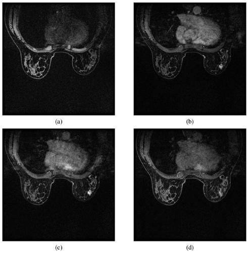
[0054] The "three-channel image" in the present invention refers to using three single-channel images (ie silhouette image, enhanced early image and enhanced late image) as one channel of the image respectively to form an image with three channels.

[0055] The mammary gland DCE-MRI image set used in the specific embodiment of the present inven...


PUM


Abstract

The invention discloses a mammary gland DCE-MRI image lesion segmentation model establishment based on hybrid convolution and segmentation method. The segmentation model establishing method comprisesthe following steps: firstly obtaining a three-channel image of each DCE-MRI sequence image in a mammary gland DCE-MRI image set, secondly constructing a mammary gland DCE-MRI image focus segmentationnetwork based on hybrid convolution and an ASPP network, and finally training the obtained segmentation network by using the three-channel images to obtain a trained segmentation model; and based onthe obtained segmentation model, preprocessing any DCE-MRI sequence image to be processed to obtain a three-channel image, and then inputting the three-channel image into the segmentation model to obtain a focus segmentation result. According to the method, 3D spatial features of the image are extracted through mixed 2D and 3D convolution, and a more accurate segmentation result is achieved; in addition, the ASPP is used for extracting multi-scale context features, so that the influence of lesion size difference on a segmentation result is effectively solved.

Description

technical field [0001] The invention belongs to the technical field of medical image analysis, and relates to a hybrid convolution-based establishment and segmentation method of a breast DCE-MRI image lesion segmentation model. Background technique [0002] With the advancement of science and technology, medical imaging technology has also made great progress, and has become one of the indispensable means of breast cancer screening, diagnosis and treatment. Magnetic Resonance Imaging (MRI) can obtain multi-angle and more comprehensive tomographic images of patients. As one of the routine screening techniques for breast diseases, MRI is of great value in the diagnosis and treatment of breast cancer. Among them, the dynamic contrast-enhanced magnetic resonance imaging (DCE-MRI) sequence has high-resolution images and dynamic information expression capabilities, and is the main sequence image for observing the internal structure and edge morphology of lesions. The segmentation...

Claims


Application Information

Patent Timeline
Patent Type & Authority Applications(China)
IPC IPC(8): G06T7/174G06T7/00G06K9/62G06N3/04G06N3/08
CPCG06T7/174G06T7/0012G06N3/08G06T2207/10016G06T2207/10096G06T2207/30068G06N3/045G06F18/253
Inventor 冯宏伟曹佳琦王红玉卜起荣冯筠
Owner NORTHWEST UNIV(CN)
Why Eureka
Try Eureka