Micro-fluidic channel, micro-fluidic chip and method for processing cells
A microfluidic channel and microfluidic chip technology, applied in the field of biomedical engineering, achieves the effects of strong controllability, good portability and fast onset
- Summary
- Abstract
- Description
- Claims
- Application Information
AI Technical Summary
Problems solved by technology
Method used
Image

Examples
Embodiment 1
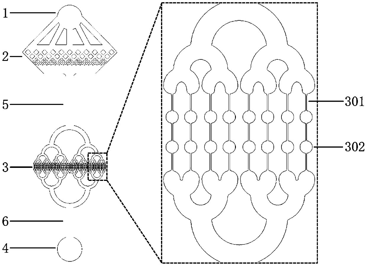
[0046] Embodiment 1 provides a microfluidic chip, and the provided microfluidic chip includes a substrate and a microfluidic channel formed on the substrate. Provided microfluidic channels such as figure 1 shown. The microfluidic channel can be divided into four parts: inlet 1, screening area 2, extrusion area 3 and outlet 4. The inlet 1 is directly connected to the screening area 2 through 5 bifurcated flow channels, and enters the extrusion area 3 after passing through the first connecting area 5 with a length of 2 mm and a width of 0.32 mm. The flow channel of the extrusion area 3 is divided into 64 extrusion The passages merge again and connect to the exit after passing through the second connecting area 6 with a length of 1.2 mm and a width of 0.32 mm. The inlet 1 and the outlet 4 are both circular with a diameter of 1 mm; the screening area 2 is arranged with rhombic columnar structures in sequence according to the direction of cell flow, and the rhombic side length of...
Embodiment 2
[0059] Embodiment 2 provides a kind of cell mechanical pre-excitation method, comprising:
[0060] 1. Prepare 3% (w / v) bovine serum albumin (BSA) solution and 0.01% (w / v) Pluronic F-127 solution in sterile phosphate buffered saline (PBS), and filter it with 0.22 μm membrane after completely dissolving filter sterilization;
[0061] 2. Use a polytetrafluoroethylene (PTFE) catheter with an outer diameter of 0.9 mm and an inner diameter of 0.5 mm to connect the syringe and the microfluidic chip (the microfluidic chip is the microfluidic chip prepared in Example 1), successively with 75% Alcohol, PBS and 3% (w / v) BSA solution degass and pre-rinse the microfluidic channel;
[0062] 3. After the mesenchymal stem cells were digested and centrifuged, they were resuspended in 0.01% (w / v) Pluronic F-127 solution to a density of 2-3×10 6 mL -1 The cell suspension was transferred to a 1mL sterile syringe;
[0063] 4. Load the syringe on the micro-injection pump, set the size of the 1 mL...
Embodiment 3
[0074] Example 3 Comparison of Cell Deformability
[0075] Referring to the design of a quantitative analyzer for cell deformability (Kenddra D.Nyberg, et al.Biophys.J., 2017, Quantitative Deformability Cytometry: Rapid, Calibrated Measurements of Cell Mechanical Properties), use AutoCAD to design microchannels for quantitative analysis of cell mechanical phenotypes (Such as Figure 6 As shown in a), this channel is to record the deformation of cells through weak transient extrusion, so as to characterize the deformability of cells (cells that have undergone this characterization are no longer suitable for use, that is, they are no longer used for culture or other applications) , the minimum line width at the narrow part has three specifications of 5, 7, and 9 μm, which can be applied to the comparison of deformability between various cells of different diameters. In this example, the mechanical phenotype quantitative analysis microfluidic channel with a line width of 5 μm wa...
PUM
| Property | Measurement | Unit |
|---|---|---|
| Outer diameter | aaaaa | aaaaa |
| The inside diameter of | aaaaa | aaaaa |
Abstract
Description
Claims
Application Information
 - R&D Engineer
- R&D Manager
- IP Professional
- Industry Leading Data Capabilities
- Powerful AI technology
- Patent DNA Extraction
Browse by: Latest US Patents, China's latest patents, Technical Efficacy Thesaurus, Application Domain, Technology Topic.
© 2024 PatSnap. All rights reserved.Legal|Privacy policy|Modern Slavery Act Transparency Statement|Sitemap