Focus analysis method and device based on medical images

A medical imaging and analysis method technology, applied in the field of image processing, can solve the problems of inaccurate recognition results, inability to analyze lesions, and only determine the location of lesions, so as to avoid overfitting, comprehensive feature information of lesions, and improve accuracy rate effect

Active Publication Date: 2020-07-31
INFERVISION MEDICAL TECH CO LTD
View PDF5 Cites 0 Cited by
  • Summary
  • Abstract
  • Description
  • Claims
  • Application Information

AI Technical Summary

Problems solved by technology

[0003] At present, in the prior art, neural networks are used to detect lesions in medical images, extract feature information of lesions, obtain lesion areas in the form of marked frames, and identify and mark lesions. Due to the use of marked frames, the specific shape of lesions It cannot be expressed, only the location of the lesion can be determined, and a more comprehensive analysis of the lesion cannot be performed
Using a

Method used


Image

  • Focus analysis method and device based on medical images
  • Focus analysis method and device based on medical images
  • Focus analysis method and device based on medical images

Examples


Embodiment Construction

[0042] The following will clearly and completely describe the technical solutions in the embodiments of the application with reference to the drawings in the embodiments of the application. Apparently, the described embodiments are only some, not all, embodiments of the application. Based on the embodiments in this application, all other embodiments obtained by persons of ordinary skill in the art without creative efforts fall within the protection scope of this application.

[0043] Application overview

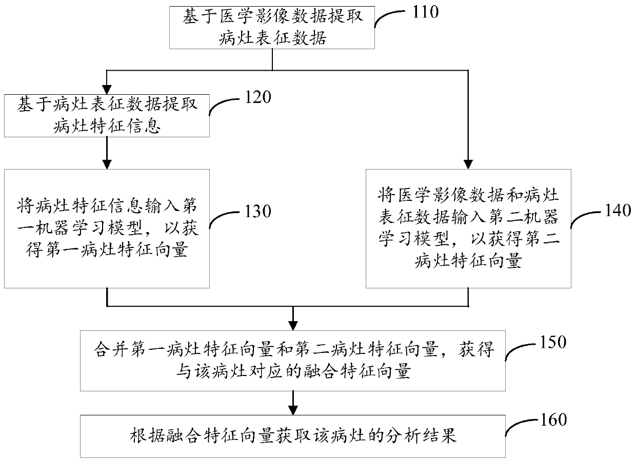
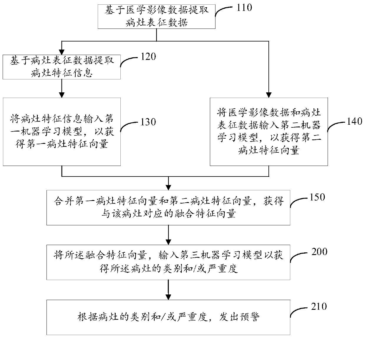
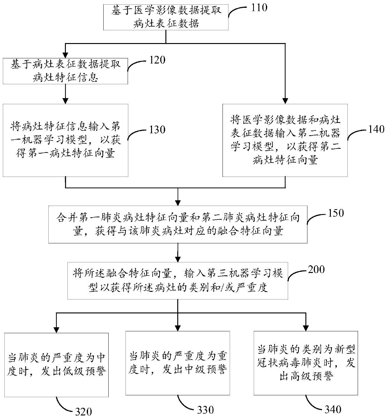
[0044] As mentioned above, in the existing analysis of lesions in medical images, only the position of the lesions is framed in the form of a marker frame, but no follow-up analysis is performed on the framed lesions, or the characteristics of the lesions in the medical images are compared with Compared with the characteristics of lesions in the database, instead of analyzing the lesions themselves in medical images, the analysis results of the lesions obtained are not com...


PUM


Abstract

The embodiment of the invention provides a focus analysis method and device based on medical images, electronic equipment and a computer readable storage medium. The invention solves the problem thatfocus analysis in the medical image is not comprehensive and not accurate enough in the prior art. The focus analysis method comprises the following steps: extracting focus characterization data basedon medical image data; extracting focus feature information based on the focus representation data; inputting the lesion feature information into a first machine learning model to obtain a first lesion feature vector; inputting the medical image data and the focus representation data into a second machine learning model to obtain a second focus feature vector; combining the first lesion feature vector and the second lesion feature vector to obtain a fusion feature vector corresponding to the lesion; and obtaining an analysis result of the focus according to the fusion feature vector.

Description

technical field [0001] The present application relates to the technical field of image processing, in particular to a method, device, electronic device and computer-readable storage medium for lesion analysis based on medical images. Background technique [0002] Pneumonia is a common respiratory inflammatory disease, mainly refers to lung infection caused by bacteria, viruses, etc. In early pneumonia, the lesion area shown on CT (Computed Tomography) images is small and not obvious. When doctors observe , it takes a lot of time to find the corresponding lesions. Recently, the novel coronavirus pneumonia is rampant, and CT images, as one of the important indicators for judging the novel coronavirus pneumonia, have greatly increased the workload of radiologists. [0003] At present, in the prior art, neural networks are used to detect lesions in medical images, extract feature information of lesions, obtain lesion areas in the form of marked frames, and identify and mark les...

Claims


Application Information

Patent Timeline
IPC IPC(8): G06T7/00G06T7/12G06T7/62G06K9/46
CPCG06T7/0012G06T7/12G06T7/62G06T2207/10081G06T2207/20084G06T2207/20081G06T2207/30061G06V10/44Y02A90/10
Inventor 尹思源张欢王瑜赵朝炜李新阳王少康陈宽
Owner INFERVISION MEDICAL TECH CO LTD
Why Eureka
Try Eureka