Fluorophore imaging devices, systems, and methods for an endoscopic procedure

An imaging device, fluorescence imaging technology, applied in endoscopy, surgery, gastroscopy, etc.

Pending Publication Date: 2020-08-14
BOSTON SCI SCIMED INC +1
View PDF5 Cites 2 Cited by
  • Summary
  • Abstract
  • Description
  • Claims
  • Application Information

AI Technical Summary

Problems solved by technology

However, contrast media is usually only used after intubation because contrast media are known catheter irritants and may put patients at risk for post-ERCP pancreatitis

Method used


Image

  • Fluorophore imaging devices, systems, and methods for an endoscopic procedure
  • Fluorophore imaging devices, systems, and methods for an endoscopic procedure
  • Fluorophore imaging devices, systems, and methods for an endoscopic procedure

Examples


Embodiment Construction

[0021] The invention is not limited to the particular embodiments described herein. The terminology used herein is for the purpose of describing particular embodiments only and is not intended to limit the scope of the appending claims. Unless otherwise defined, all technical terms used herein have the same meaning as commonly understood by one of ordinary skill in the art to which this invention belongs.

[0022] As used herein, the singular forms "a", "an" and "the" are intended to include the plural forms as well, unless the content clearly dictates otherwise. It will be further understood that when used herein, the term "comprises" and / or "comprises", or "includes" and / or "comprises", designates said features, regions, step elements and / or components but does not preclude the presence or addition of one or more other features, regions, integers, steps, operations, elements, components and / or groups thereof.

[0023] The devices, systems and methods described herein aim t...


PUM

PropertyMeasurementUnit
optical densityaaaaaaaaaa
transmittivityaaaaaaaaaa

Abstract

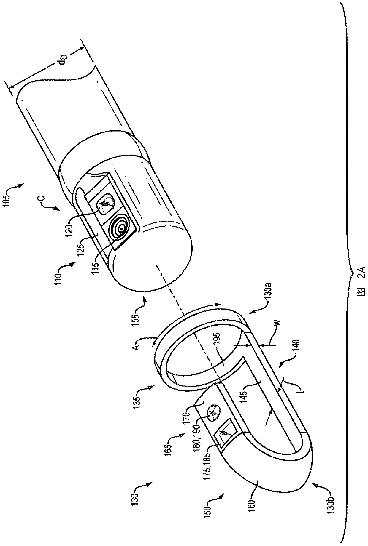
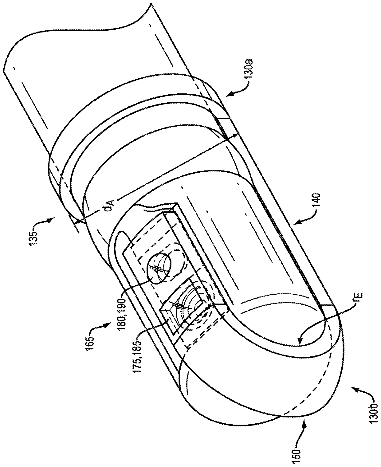
Fluorescent imaging systems for performing an endoscopic procedure, such as a retrograde cholangiopancreatography (ERCP) procedure may include a first light source for emitting light in the visible spectrum, or light in the near infrared (NIR) spectrum, or both. A light source bandpass filter may block the emitted light in the visible spectrum, or in the NIR spectrum, or both. A first sensor may be capable of detecting the light in the visible spectrum, or the light in the NIR spectrum, or both. A sensor bandpass filter may block the detected light in the visible spectrum, or in the NIR spectrum, or both. The first or a second light source, or the first or a second sensor, or combinations thereof, may be removably disposed on a duodenoscope.

Description

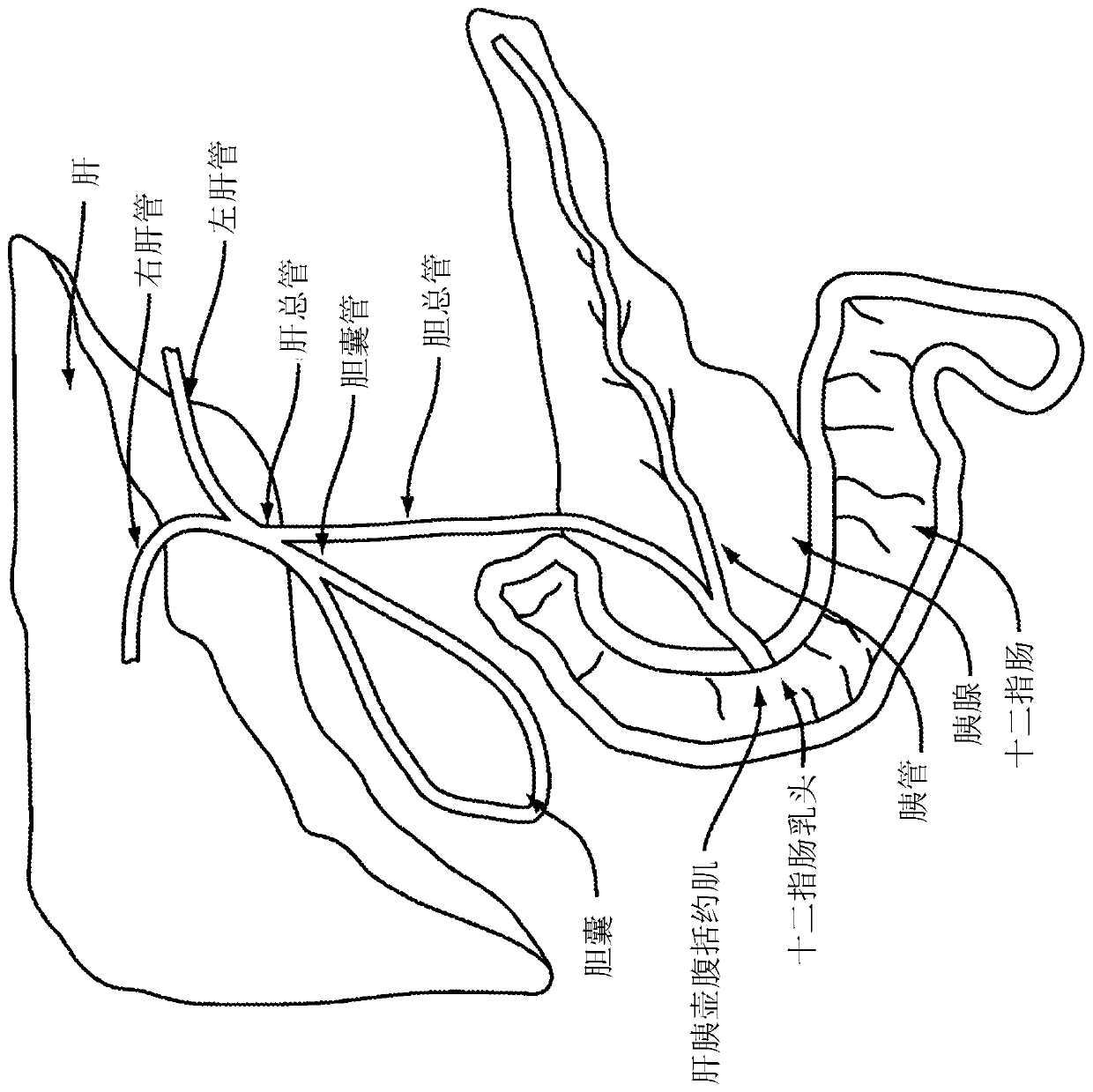
[0001] Cross References to Related Applications [0002] This application claims priority to U.S. Provisional Patent Application No. 62 / 614,266, entitled "Fluorophore Imaging Apparatus, System, and Method for Endoscopic Surgery," filed January 5, 2018, which is incorporated by reference It is hereby incorporated in its entirety. technical field [0003] The present invention relates generally to fluorographic imaging devices, systems, and methods for endoscopic surgery, and more particularly, to the use of indocyanine green (ICG) for the use of indocyanine green (ICG) during endoscopic retrograde cholangiopancreatography (ERCP). The bile ducts of the duodenum are imaged. Background technique [0004] During endoscopic procedures, such as ERCP procedures, selective cannulation provides access through the duodenal papilla into the patient's bile duct or pancreatic duct. Medical personnel may not easily see the direction of the catheter and may incorrectly position and / or or...

Claims


Application Information

Patent Timeline
Patent Type & Authority Applications(China)
IPC IPC(8): A61B1/04A61B1/00A61B1/018A61B1/273
CPCA61B1/00096A61B1/00101A61B1/00137A61B1/00177A61B1/018A61B1/2736A61B1/00186A61B1/043A61B1/00165A61B1/05A61B1/0646A61B1/0661A61B1/07
Inventor 杰西卡·鲍尔斯雅各布·考德威尔晟敏·R·曹尤斯菲·雅兹迪尼古拉斯·J·杜尔潘卡耶·J·帕斯理查查德·维勒阿希什·尼姆冈卡尔
Owner BOSTON SCI SCIMED INC
Why Eureka
Try Eureka