Three-dimensional craniofacial image feature point marking analysis system and method

An analysis system and feature point technology, applied in image analysis, neural learning methods, echo tomography, etc., can solve the problems of increasing the positioning time of craniofacial anatomical feature points, loss of details, inability to in-depth reasoning and evaluation, etc., to achieve The effect of reducing the risk of wrong diagnosis and improving the overall efficiency

Pending Publication Date: 2020-08-28
SHANGHAI UEG MEDICAL IMAGING EQUIP CO LTD
View PDF17 Cites 9 Cited by
  • Summary
  • Abstract
  • Description
  • Claims
  • Application Information

AI Technical Summary

Problems solved by technology

[0002] Three-dimensional craniofacial image analysis technology is a method to evaluate the overall shape and function of the patient's craniofacial image by analyzing the CT images of the patient's craniofacial region. In traditional methods, CBCT imaging equipment can only complete the three-dimensional craniofacial model of the patient. reconstruction, but cannot carry out more in-depth reasoning and evaluation. In the specific operation, after the reconstruction of the 3D model is completed, it is necessary to analyze according to the reconstruction results, and generate a me

Method used


Image

  • Three-dimensional craniofacial image feature point marking analysis system and method
  • Three-dimensional craniofacial image feature point marking analysis system and method
  • Three-dimensional craniofacial image feature point marking analysis system and method

Examples


Embodiment Construction

[0057] In order to make the technical means, creative features, objectives and effects of the invention easy to understand, the present invention will be further elaborated below in conjunction with specific illustrations.

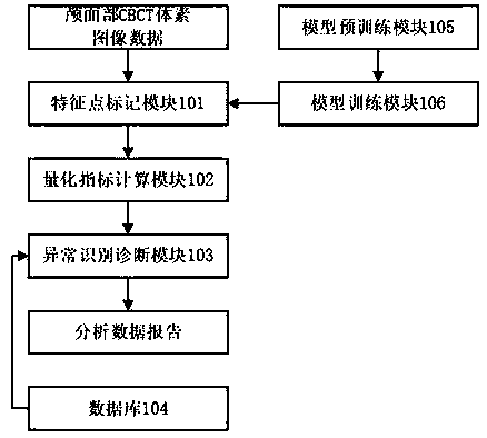
[0058] The first embodiment of the present invention is to provide a three-dimensional craniofacial image feature point marker analysis system, the purpose is that after the image equipment completes the craniofacial three-dimensional image reconstruction of the image acquisition object, the present invention can perform anatomical feature point analysis on the three-dimensional image Identification; then the analysis method described in this patent will automatically analyze the cranial face according to the identified feature point information, and output the analysis result in the form of an analysis report. The feature point recognition result and analysis report can be used as the basis for the doctor's subsequent diagnosis. Significantly reduce the wo...


PUM


Abstract

The invention discloses a three-dimensional craniofacial image feature point marking analysis system and method. The system comprises a feature point marking module, a quantitative index calculation module, an anomaly recognition and diagnosis module and a database. The feature point marking module is used for acquiring craniofacial CBCT voxel image data and marking craniofacial anatomical structure feature points according to the craniofacial CBCT voxel image data to generate feature point coordinates; the quantitative index calculation module is used for acquiring coordinates of the featurepoints and calculating quantitative index data according to the coordinates of the feature points; the anomaly recognition and diagnosis module is used for acquiring the coordinates of the feature points, quantifying index data and comparing the quantified index data with sample data in a database to generate an analysis data report. The method comprises the following steps: marking the craniofacial CBCT voxel image data to obtain the feature point coordinates; calculating the coordinates of the feature points to obtain the quantitative index data; and comparing the feature point coordinates and the quantitative index data with sample data in the database to generate the analysis data report.

Description

technical field [0001] The invention relates to the technical field of computer-aided diagnosis, in particular to a system and method for marking and analyzing feature points of three-dimensional craniofacial images. Background technique [0002] Three-dimensional craniofacial image analysis technology is a method to evaluate the overall shape and function of the patient's craniofacial image by analyzing the CT images of the patient's craniofacial region. In traditional methods, CBCT imaging equipment can only complete the three-dimensional craniofacial model of the patient. reconstruction, but cannot carry out more in-depth reasoning and evaluation. In the specific operation, after the reconstruction of the 3D model is completed, it is necessary to analyze according to the reconstruction results, and generate a medical diagnosis report according to the usage scenario to assist doctors in diagnosis and treatment; [0003] CBCT is the abbreviation of Cone beam CT, that is, ...

Claims


Application Information

Patent Timeline
IPC IPC(8): G16H15/00G16H50/20G06T17/00G06T7/00G06N3/08G06N3/04G06K9/62G06K9/00A61B6/03A61B6/00
CPCG16H15/00G16H50/20G06N3/08A61B6/032A61B6/4085A61B6/5211A61B6/52A61B6/5294G06T17/00G06T7/0012G06T2207/10116G06T2207/10012G06T2207/30204G06V40/169G06N3/045G06F18/253G06F18/214
Inventor 杜鑫陈毅朱露张彬琳
Owner SHANGHAI UEG MEDICAL IMAGING EQUIP CO LTD
Why Eureka
Try Eureka