Retinal neovascularization detection method and imaging method for color fundus image

A fundus image and new blood vessel technology, applied in the field of image processing, can solve the problems of difficult effective features and increase the complexity of the method, and achieve the effect of simple detection and imaging, good practicability and high reliability

Active Publication Date: 2020-09-01
CENT SOUTH UNIV
View PDF7 Cites 3 Cited by
  • Summary
  • Abstract
  • Description
  • Claims
  • Application Information

AI Technical Summary

Problems solved by technology

However, the extraction of effective features is a difficult process, and such methods also need to segment retinal vessels first, which further increases the complexity of the method

Method used


Image

  • Retinal neovascularization detection method and imaging method for color fundus image
  • Retinal neovascularization detection method and imaging method for color fundus image
  • Retinal neovascularization detection method and imaging method for color fundus image

Examples


Embodiment Construction

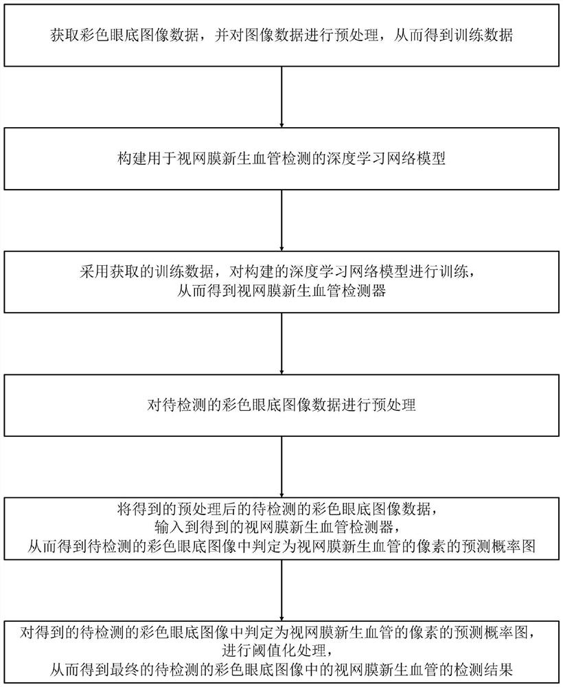
[0067] Such as figure 1 Shown is a schematic flow chart of the detection method of the present invention: the retinal neovascularization detection method for color fundus images provided by the present invention includes the following steps:

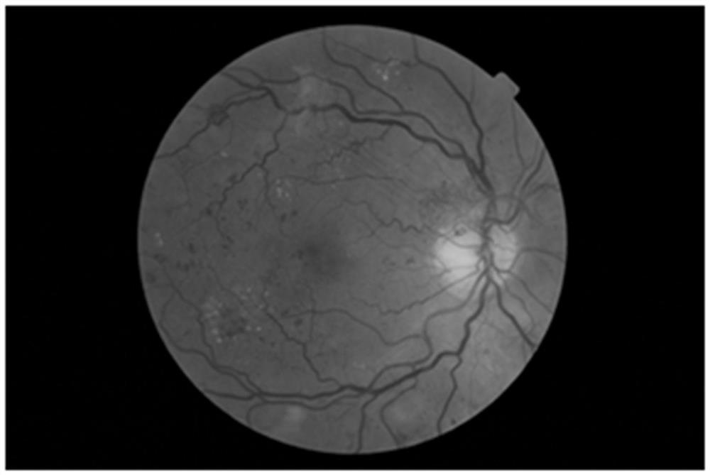
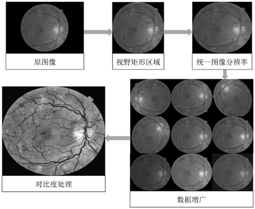
[0068] S1. Obtain color fundus image data (such as figure 2 As shown), and preprocess the image data to obtain the training data; because the resolution of the image and the size of the field of view are different, it will affect the mining of retinal neovascularization features in the model detection process, so it is necessary to perform a series of images on the image first. The preprocessing operation; Specifically, the following steps are adopted (such as image 3 shown) for preprocessing:

[0069] A. For the image data, cut out the field of view area; specifically: first convert the color fundus image data into a grayscale image; then perform threshold processing on the grayscale image, thereby converting the grayscale image int...


PUM


Abstract

The invention discloses a retinal neovascularization detection method for a color fundus image. The method comprises the steps of obtaining color fundus image data and performing preprocessing to obtain training data; constructing a deep learning network model for retinal neovascularization detection; training the deep learning network model by adopting the training data to obtain a retinal neovascularization detector; preprocessing the color fundus image data to be detected; adopting a retinal neovascularization detector to detect the preprocessed to-be-detected color eye fundus image data toobtain a prediction probability graph of pixels judged as retinal neovascularization in the to-be-detected color eye fundus image; and carrying out thresholding processing to obtain a final detectionresult of the retinal neovascularization in the to-be-detected color fundus image. The invention further discloses an imaging method comprising the retinal neovascularization detection method for thecolor fundus picture. The device is high in reliability, good in practicability and good in detection effect.

Description

technical field [0001] The invention belongs to the field of image processing, and in particular relates to a method for detecting retinal neovascularization and an imaging method for color fundus images. Background technique [0002] With the development of economy and technology and the improvement of people's living standards, people are paying more and more attention to eye health. Retinal neovascularization is an important reflection of the state of the eye and has high clinical application value. Therefore, the detection of retinal neovascularization has always been the focus of research. [0003] At present, for the detection of retinal neovascularization, eye fluorescein angiography or color fundus image detection is generally used. Although ocular fluorescein angiography can clearly and accurately reflect the relevant information of the examiner's retinal neovascularization, it is an invasive test, and the test results are greatly affected by time factors, so the ...

Claims


Application Information

Patent Timeline
Patent Type & Authority Applications(China)
IPC IPC(8): G06T7/90G06T7/136G06T7/11G06T7/00G06T5/50G06T5/40G06T3/40G06N3/04G06N3/08
CPCG06T7/0012G06T7/11G06T3/4023G06T5/40G06T7/136G06T5/50G06T7/90G06N3/08G06T2207/20016G06T2207/20081G06T2207/20132G06T2207/20221G06T2207/30041G06T2207/30101G06N3/045Y02T10/40
Inventor 邹北骥戴玉兰朱承璋欧阳平波刘耕许利群陈庆勇
Owner CENT SOUTH UNIV
Why Eureka
Try Eureka