A method for evaluating fundus image quality

A fundus image and image technology, applied in the field of medical image processing, can solve problems such as low contrast, insufficient or too strong light, and blur

Active Publication Date: 2020-09-08
宁波蓝明信息科技有限公司
View PDF8 Cites 5 Cited by
  • Summary
  • Abstract
  • Description
  • Claims
  • Application Information

AI Technical Summary

Problems solved by technology

This method evaluates the quality of fundus images weightedly for the reasons that easily lead to low f

Method used


Image

  • A method for evaluating fundus image quality

Examples


Embodiment Construction

[0008] Specific embodiments of the present invention will be described in detail below in combination with technical solutions and accompanying drawings.

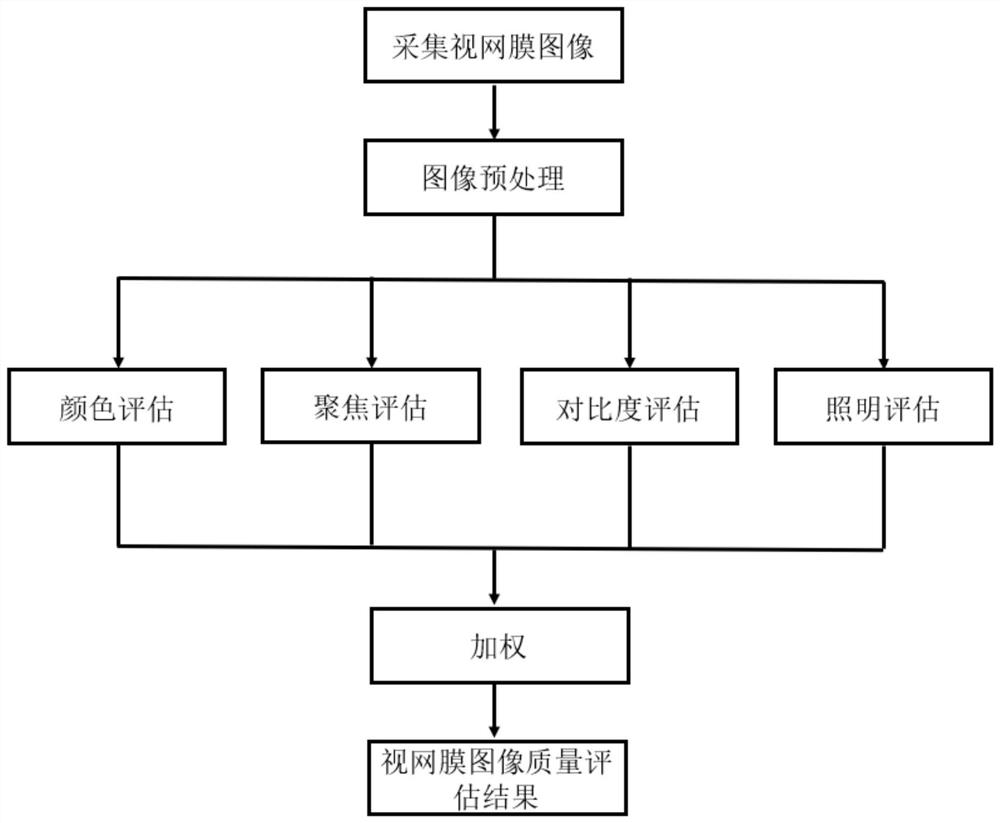
[0009] A method for evaluating the quality of fundus images, specifically comprising the steps of:

[0010] Step 1, preprocessing the fundus image of the subject collected by the fundus camera, cutting out the redundant background around the fundus image, and obtaining the area containing only the retina;

[0011] In this implementation example, step 1 converts the retinal image into a grayscale image. And using the threshold segmentation algorithm to get the mask image, in the foreground area with the center point of the image as the center, find the largest radius to make a circular area, the circular area includes all the foreground area, and finally use the circular area to cut out the retinal image.

[0012] Step 2, based on the preprocessed fundus image, extract and evaluate color, focus, contrast and illumination fe...


PUM


Abstract

The invention belongs to the field of medical image processing, and discloses a method for evaluating fundus image quality. The method comprises the following steps: preprocessing an acquired fundus image, and cutting redundant backgrounds around a retina image to obtain an area only containing a retina; then, respectively extracting color, focusing, contrast and illumination features based on thepreprocessed fundus image, and evaluating the color, focusing, contrast and illumination features; and finally, determining an evaluation result of the fundus image based on the confidence coefficient of the feature weighted evaluation, and analyzing a reason causing low imaging quality. Therefore, the method can be used for guiding the retinal image acquisition process, the workload of ophthalmologists in screening is reduced, and the cost effectiveness is improved.

Description

technical field [0001] The invention relates to the field of medical image processing, in particular to a method for evaluating the quality of fundus images. Background technique [0002] With the development of my country's social economy and the change of people's diet and living habits, the incidence of diabetes is increasing year by year. At present, my country has become the country with the largest number of diabetic patients in the world. Among them, diabetic retinopathy is one of the common complications of diabetes, and its incidence rate is as high as more than 40% in diabetic patients. Diabetic retinopathy has become one of the main causes of vision loss and even blindness. However, through early diagnosis and treatment of diabetic retinopathy patients, visual impairment and blindness can be prevented in more than 50% of patients. Therefore, fundus screening is crucial for the early diagnosis of diabetic retinopathy and the prevention of blindness. [0003] Howe...

Claims


Application Information

Patent Timeline
IPC IPC(8): G06T7/00G06T7/11G06T7/194
CPCG06T7/0002G06T7/11G06T7/194G06T7/0012G06T2207/30168G06T2207/30041G06T2207/20132G06T2207/10024G06T2207/30101G06T2207/20056
Inventor 孔令敏陈硕佟萌萌朱姗姗
Owner 宁波蓝明信息科技有限公司
Why Eureka
Try Eureka