CT image separation and reconstruction method and application

A CT image and image reconstruction technology, applied in the field of medical CT imaging, can solve problems such as X-ray radiation hazards, and achieve the effect of artifact elimination

Active Publication Date: 2020-09-25
NAT INST OF ADVANCED MEDICAL DEVICES SHENZHEN
View PDF5 Cites 0 Cited by
  • Summary
  • Abstract
  • Description
  • Claims
  • Application Information

AI Technical Summary

Problems solved by technology

[0006] Based on the existing dynamic perfusion CT image reconstruction method, image reconstruction can only be performed based on fully sampled scan data. In the process of repeated scanning of the same part of the patient, there is a pheno

Method used


Image

  • CT image separation and reconstruction method and application
  • CT image separation and reconstruction method and application
  • CT image separation and reconstruction method and application

Examples


Embodiment Construction

[0030] Hereinafter, specific embodiments of the present application will be described in detail with reference to the accompanying drawings. According to these detailed descriptions, those skilled in the art can clearly understand the present application and can implement the present application. Without departing from the principle of the present application, the features in different embodiments can be combined to obtain new implementations, or some features in certain embodiments can be replaced to obtain other preferred implementations.

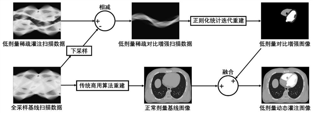
[0031] Clinical myocardial dynamic perfusion CT imaging includes baseline scanning before the imaging agent reaches the heart tissue (including aorta, coronary artery, left and right ventricle, left and right atrium, and myocardial tissue) and imaging agent after the imaging agent reaches the heart tissue. Continuous dynamic perfusion scan. Under the existing clinical conditions, the baseline scan and the dynamic perfusion scan adopt the ...


PUM


Abstract

The invention belongs to the technical field of medical CT imaging, and particularly relates to a CT image separation and reconstruction method and application. A sparse-angle X-ray scanning scheme can significantly reduce the radiation dose caused by repeated scanning, but there is no effective and feasible method for image reconstruction of sparse-angle image acquisition data. The invention provides a CT image separation and reconstruction method. The method comprises the following steps: step 1, separating a contrast-enhanced scanning image introduced by a developer from a dynamic perfusionscanning image; 2, reconstructing an enhanced image by using the contrast enhanced scanning image; and 3, fusing the enhanced image with a baseline image obtained by reconstructing a full-sampling baseline image acquisition image to obtain a perfusion CT reconstructed image. And artifacts in the fused dynamic perfusion image can be eliminated to a great extent.

Description

technical field [0001] The application belongs to the technical field of medical CT imaging, and in particular relates to a separation and reconstruction method and application of CT images. Background technique [0002] CT is a full-featured disease detection instrument, which is the abbreviation of computerized X-ray tomography. The scanner used in computed axial tomography (CAT) produces X-rays, a powerful form of electromagnetic energy. X-ray photons are basically the same as photons of ordinary visible light, but they carry more energy. This higher energy level allows X-rays to pass directly through most of the body's soft tissues (see X-Ray Introduction to learn how X-rays penetrate soft tissue and how X-ray machines generate X-ray photons). Conventional X-ray imaging technology utilizes the principle of light and shadow. When "light" is shone from one side of the body, the film on the other side of the body records the outline of the bones. [0003] Myocardial per...

Claims


Application Information

Patent Timeline
IPC IPC(8): G06T11/00
CPCG06T11/003G06T2210/41G06T2211/416
Inventor 郑海荣李彦明万丽雯胡战利陈子翔
Owner NAT INST OF ADVANCED MEDICAL DEVICES SHENZHEN
Why Eureka
Try Eureka