Imaging method and device for self-luminous object on biological sample film

A biological sample and imaging device technology, applied in the field of sample analysis, can solve the problems of complex operation process, high cost, and time-consuming imaging, and achieve the effects of simple operation process, low manufacturing cost, and short imaging time-consuming

Pending Publication Date: 2020-10-09
SANGHAI E BLOT PHOTOELECTRIC TECHNOLOGY CO LTD
View PDF0 Cites 2 Cited by
  • Summary
  • Abstract
  • Description
  • Claims
  • Application Information

AI Technical Summary

Problems solved by technology

[0004] The technical problem to be solved by the present invention is that the imaging method for obtaining self-luminous objects on the biological sample film in the prior art has complex operation process, long imaging time, and the imaging system has large volume, large space occupation, high cost, and is not easy to transport. and other defects, the purpose is to provide an imaging method and device for self-luminous objects on biological sample films

Method used


Image

  • Imaging method and device for self-luminous object on biological sample film
  • Imaging method and device for self-luminous object on biological sample film
  • Imaging method and device for self-luminous object on biological sample film

Examples


Embodiment 1

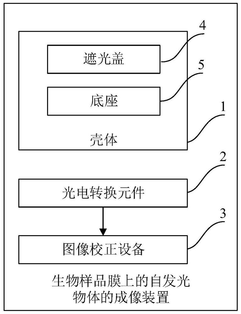
[0078] Such as figure 1 and figure 2 As shown, the imaging device for a self-luminous object on a biological sample film in this embodiment includes a casing 1 , a photoelectric conversion element 2 and an image correction device 3 .

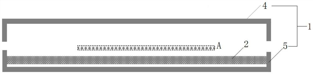
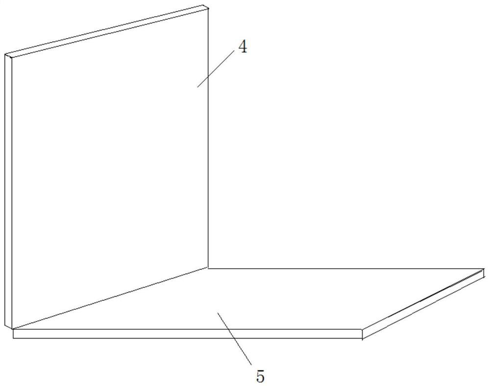
[0079] The interior of the housing 1 constitutes a darkroom space, wherein the housing 1 includes a light-shielding cover 4 and a base 5, such as image 3 As shown, one side of the shade cover 4 is hinged to one side of the base 5, and the shade cover 4 is in an open state at this moment.

[0080] Such as Figure 4 As shown, when the light-shielding cover 4 and the base 5 are closed, the interior of the housing 1 forms a darkroom space to ensure the biological sample film (in figure 2 The weak signal corresponding to the self-emitting object on the surface is represented by A) to effectively collect.

[0081] Wherein, biological sample membranes include protein membranes, agarose gel blocks, agarose gel strips, polyacrylamide gel gel block...

Embodiment 2

[0103] Such as Figure 7 As shown, the imaging device of the self-luminous object on the biological sample film of this embodiment is a further improvement to Embodiment 1, specifically:

[0104] The imaging device further includes a light source device 6 , and the light source device 6 is arranged in the casing 1 .

[0105] The light source device 6 is arranged at the top position inside the light shielding cover 4;

[0106] The corresponding illumination duration after the light source device 6 is turned on is 10ms-30s, and the illumination duration can also be adjusted according to actual needs.

[0107] The light source device 6 includes a number of LED lamp beads arranged in a dot matrix, a number of lights guided by optical fibers, a number of parallel arranged light tubes, a number of plate-shaped lights, and the like.

[0108] The imaging device also includes a diffuser plate 7 , which is fixed inside the light-shielding cover and directly below the light source devi...

Embodiment 3

[0136] The imaging device of the self-luminous object on the biological sample film of this embodiment is a further improvement on Embodiment 1, specifically:

[0137] This embodiment includes the light source device 6 in Embodiment 2.

[0138] The photoelectric conversion element 2 is also used to obtain bright field images corresponding to the set acquisition time period when the biological sample film is not placed in the housing and a uniform light field is provided by an external light source, that is, the first dark field image, the second dark field image, and the second dark field image. The data acquisition time corresponding to the image and the bright field image are the same;

[0139] The image correction device 3 is also used to correct the second dark field image according to the first dark field image and the bright field image, and obtain the target image corresponding to the self-luminous object, such as Figure 10 As shown, the difference between this pictur...


PUM

PropertyMeasurementUnit
thicknessaaaaaaaaaa

Abstract

The invention discloses an imaging method and device for a self-luminous object on a biological sample film. The imaging device comprises a shell, a photoelectric conversion element and image correction equipment, a darkroom space is formed in the shell; the photoelectric conversion element is arranged in the shell; the photoelectric conversion element is used for acquiring a first dark field image inside the shell when the biological sample film is not placed; after the biological sample film is attached to the surface of the photoelectric conversion element, the photoelectric conversion element is also used for acquiring a second dark field image in the shell; and the image correction equipment is used for correcting the second dark field image according to the first dark field image toobtain a target image corresponding to the self-luminous object. According to the invention, the target image of the self-luminous object with higher definition can be effectively and more accuratelyacquired, the operation process is simple, the imaging time is shorter, and the imaging device has the advantages of small structure, low manufacturing cost, convenience in operation, convenience in carrying and the like.

Description

technical field [0001] The invention relates to the technical field of sample analysis, in particular to an imaging method and device for a self-luminous object on a biological sample film. Background technique [0002] Currently, there are two types of biological self-luminescence detection technologies commonly used, one is to use photosensitive film technology, and the other is to use camera shooting technology. [0003] Among them, the photosensitive film technology is similar to the traditional method of developing photographic film. In the darkroom, the self-luminous object (attached to the biological sample film) is closely attached to the photosensitive film, and then the photosensitive film is rewashed with a developer. In order to obtain the image corresponding to the self-luminous object; however, this imaging method has a complicated operation process and requires more steps for digital processing; the camera shooting technology is similar to that of a mobile pho...

Claims


Application Information

Patent Timeline
Patent Type & Authority Applications(China)
IPC IPC(8): G01N21/63
CPCG01N21/63G01N21/76G01N21/274G02B21/10G02B21/365
Inventor 张英豪奚岩
Owner SANGHAI E BLOT PHOTOELECTRIC TECHNOLOGY CO LTD
Why Eureka
Try Eureka