Standard section detection method of uterus three-dimensional ultrasonic image

A technology of three-dimensional ultrasound and detection method, applied in the field of ultrasound, can solve problems such as affecting the accuracy of uterus-related conditions, and achieve the effects of realizing automatic acquisition, avoiding dependence on experience, and improving the accuracy of judgment.

Pending Publication Date: 2020-10-13
深圳度影医疗科技有限公司
View PDF0 Cites 5 Cited by
  • Summary
  • Abstract
  • Description
  • Claims
  • Application Information

AI Technical Summary

Problems solved by technology

However, when the goal is to determine the standard view in the 3D ultrasound volume data, the doctor needs to manually calibrate the standard view, which is extremely dependent on the doctor's experience and professional level, so that there are differences between the standard views obtained by different doctors, which will affect the uterus. Accuracy of Judgment about the Circumstances

Method used


Image

  • Standard section detection method of uterus three-dimensional ultrasonic image
  • Standard section detection method of uterus three-dimensional ultrasonic image
  • Standard section detection method of uterus three-dimensional ultrasonic image

Examples


Embodiment Construction

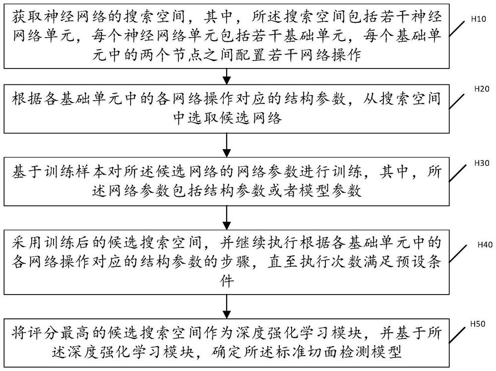
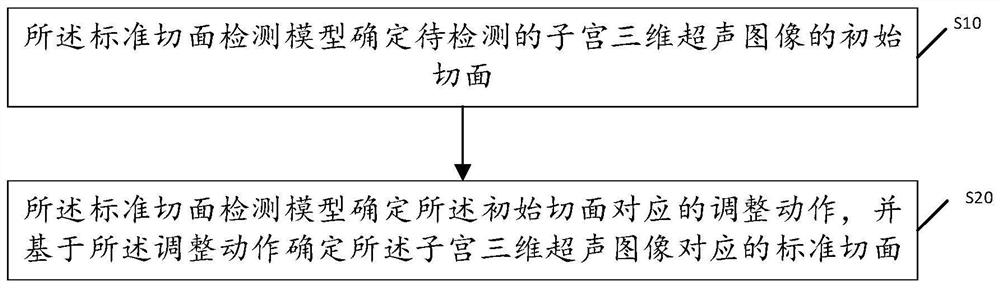
[0057] The present invention provides a standard slice detection method of a three-dimensional ultrasound image of the uterus. In order to make the purpose, technical solution and effect of the present invention clearer and clearer, the present invention will be further described in detail below with reference to the accompanying drawings and examples. It should be understood that the specific embodiments described here are only used to explain the present invention, not to limit the present invention.

[0058] Those skilled in the art will understand that unless otherwise stated, the singular forms "a", "an", "said" and "the" used herein may also include plural forms. It should be further understood that the word "comprising" used in the description of the present invention refers to the presence of said features, integers, steps, operations, elements and / or components, but does not exclude the presence or addition of one or more other features, Integers, steps, operations, e...


PUM


Abstract

The invention discloses a standard section detection method of a uterus three-dimensional ultrasonic image, which is applied to a standard section detection model, and comprises the following steps: the standard section detection model determines an initial section of the uterus three-dimensional ultrasonic image to be detected; and the standard section detection model determines an adjustment action corresponding to the initial section, and determines a standard section corresponding to the uterus three-dimensional ultrasonic image based on the adjustment action. According to the invention, the standard section corresponding to the uterus three-dimensional ultrasonic image is automatically determined through the standard section detection model; the automatic acquisition of the standard sections is realized, and the dependence of the standard sections on the experience of doctors is avoided, so that the existence of the standard sections acquired by different doctors is avoided, and the judgment accuracy of the uterus related conditions based on the standard sections is improved.

Description

technical field [0001] The invention relates to the field of ultrasound technology, in particular to a method for detecting a standard slice of a three-dimensional ultrasound image of the uterus. Background technique [0002] There are multiple standard slices in the three-dimensional ultrasound volume data of the uterus, which can be used to evaluate various abnormalities of the uterus and measure the biomass, etc. Measurement and judgment of endometrial typing, etc.), so the standard section detection of uterine three-dimensional ultrasound volume data has a very important role in clinical practice. However, when the goal is to determine the standard view in the 3D ultrasound volume data, the doctor needs to manually calibrate the standard view, which is extremely dependent on the doctor's experience and professional level, so that there are differences between the standard views obtained by different doctors, which will affect the uterus. The accuracy of judgments about ...

Claims


Application Information

Patent Timeline
Patent Type & Authority Applications(China)
IPC IPC(8): G06T7/00G06N3/04A61B5/00
CPCG06T7/0012A61B5/4325A61B5/4337G06T2207/10132G06T2207/20081G06T2207/20084G06N3/045
Inventor 杨鑫熊奕易艳高睿黄雨灏
Owner 深圳度影医疗科技有限公司
Why Eureka
Try Eureka