Choroidal vessel lumen area recognition method, device and equipment and medium

A region identification and choroid technology, applied in image data processing, instruments, calculations, etc., can solve the problems of high doctor experience requirements, manual identification deviation, low resolution of acquisition equipment, etc., to improve the accuracy and reliability of identification, reduce The effect of manual identification cost

Pending Publication Date: 2020-11-06
PING AN TECH (SHENZHEN) CO LTD
View PDF4 Cites 4 Cited by
  • Summary
  • Abstract
  • Description
  • Claims
  • Application Information

AI Technical Summary

Problems solved by technology

[0003] In the medical field, doctors often need to manually identify the luminal area of ​​the fundus choroid vessels in the collected fundus photos by experience to determine the characteristics of the fundus choroid, and then perform other medical actions based on the identified features. Ther...

Method used


Image

  • Choroidal vessel lumen area recognition method, device and equipment and medium
  • Choroidal vessel lumen area recognition method, device and equipment and medium
  • Choroidal vessel lumen area recognition method, device and equipment and medium

Examples


Embodiment Construction

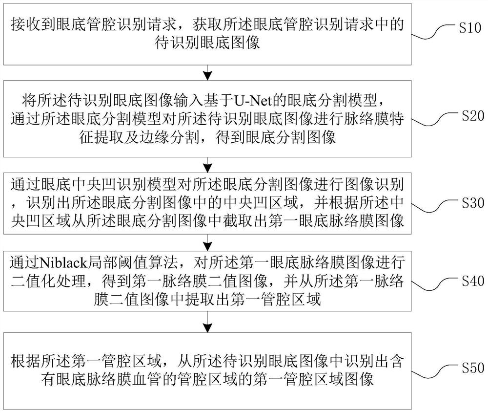
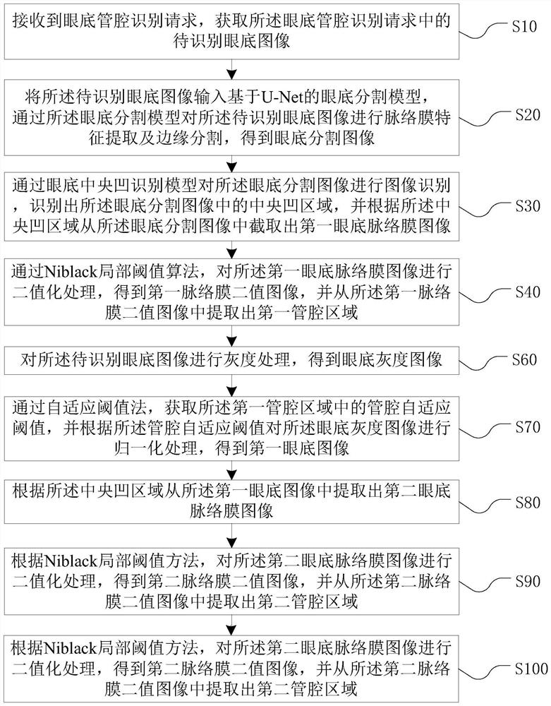
[0030] The following will clearly and completely describe the technical solutions in the embodiments of the present invention with reference to the accompanying drawings in the embodiments of the present invention. Obviously, the described embodiments are some of the embodiments of the present invention, but not all of them. Based on the embodiments of the present invention, all other embodiments obtained by persons of ordinary skill in the art without creative efforts fall within the protection scope of the present invention.

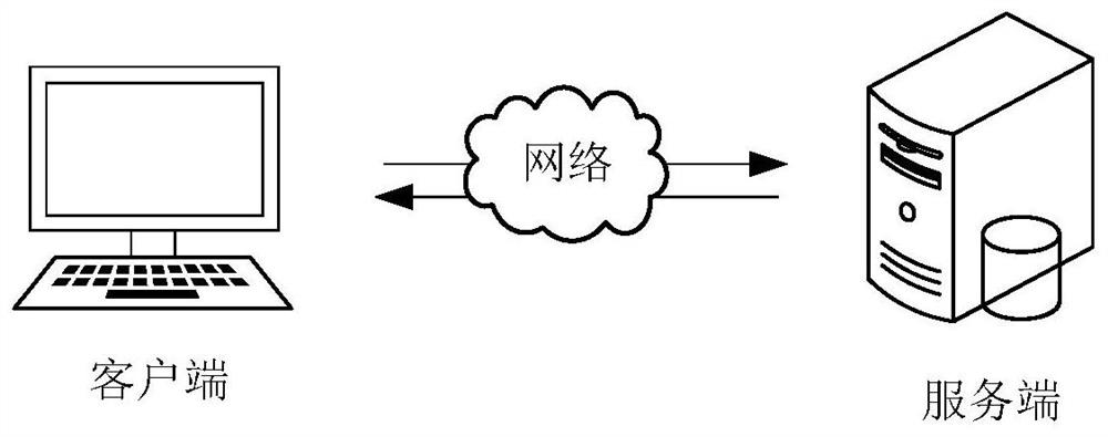
[0031] The luminal area identification method of choroidal vessels provided by the present invention can be applied in such as figure 1 , where a client (computer device) communicates with a server over a network. Wherein, the client (computer device) includes but is not limited to various personal computers, notebook computers, smart phones, tablet computers, cameras and portable wearable devices. The server can be implemented by an independent serve...


PUM


Abstract

The invention relates to the field of artificial intelligence, and provides a choroidal vessel lumen area recognition method, device and equipment and a medium. The method comprises the steps of obtaining a fundus image to be recognized; inputting a fundus segmentation model based on U-Net, and performing choroidal feature extraction and edge segmentation on the fundus image to be recognized through the fundus segmentation model to obtain a fundus segmentation image; recognizing a foveal area in the fundus segmentation image through a fundus foveal recognition model, and intercepting a first fundus choroid image from the fundus segmentation image according to the foveal area; carrying out binarization processing on the first fundus choroidal image through a Niback local threshold algorithmto obtain a first choroidal binary image, and extracting a first lumen area from the first choroidal binary image; and identifying the first lumen region image. According to the invention, the funduschoroidal vessel lumen area in the fundus image can be automatically identified. The invention is suitable for the field of smart medical treatment, and can further promote the construction of smartcities.

Description

technical field [0001] The invention relates to the field of image processing of artificial intelligence, in particular to a method, device, equipment and medium for identifying a lumen region of a choroidal vessel. Background technique [0002] The fundus choroid is located between the retina and the sclera. It is a soft, smooth, elastic, and rich-vascular brown film. It starts from the serrated edge at the front and ends around the optic nerve at the back; The pigment epithelium is connected, and the outside is connected to the sclera through a potential gap. The fine fibrous platelets of the choroidal layer are stretched and mixed into the brown and black plate of the sclera, and blood vessels and nerves pass through it. The choroid is mainly composed of blood vessels that supply oxygen and blood to the retina. [0003] In the medical field, doctors often need to manually identify the luminal area of ​​the fundus choroid vessels in the collected fundus photos by experien...

Claims


Application Information

Patent Timeline
IPC IPC(8): G06T7/00G06T7/12G06T7/136G06T7/62
CPCG06T7/0012G06T7/12G06T7/136G06T7/62G06T2207/10101G06T2207/20081G06T2207/20084G06T2207/30041G06T2207/30101
Inventor 周侠王玥张成奋吕彬吕传峰
Owner PING AN TECH (SHENZHEN) CO LTD
Why Eureka
Try Eureka