
Brain image segmentation method and device, computer equipment and readable storage medium

A computer program and brain technology, applied in the field of image processing, can solve the problem of low accuracy of intracranial hemorrhage segmentation results, and achieve the effect of improving accuracy

Pending Publication Date: 2020-11-17
SHANGHAI UNITED IMAGING INTELLIGENT MEDICAL TECH CO LTD
View PDF0 Cites 3 Cited by
  • Summary
  • Abstract
  • Description
  • Claims
  • Application Information

AI Technical Summary

Problems solved by technology

[0005] Based on this, it is necessary to provide a brain image segmentation method, device, computer equipment and readable storage medium for the problem of low accuracy of intracranial hemorrhage segmentation results obtained by deep learning networks in traditional technologies

Method used


Image

  • Brain image segmentation method and device, computer equipment and readable storage medium
  • Brain image segmentation method and device, computer equipment and readable storage medium
  • Brain image segmentation method and device, computer equipment and readable storage medium

Examples


Embodiment Construction

[0047] In order to make the purpose, technical solution and advantages of the present application clearer, the present application will be further described in detail below in conjunction with the accompanying drawings and embodiments. It should be understood that the specific embodiments described here are only used to explain the present application, and are not intended to limit the present application.

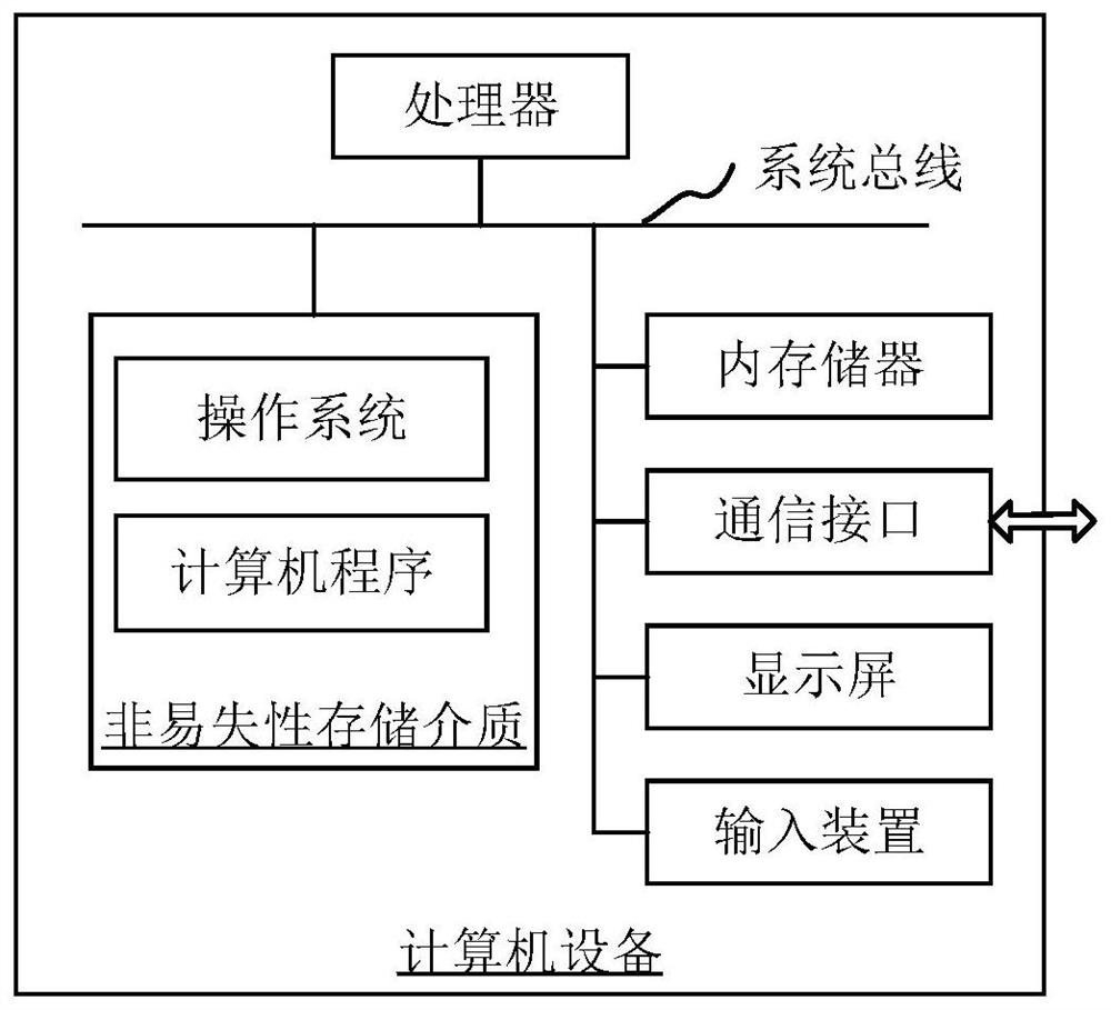
[0048] The brain image segmentation method provided by the embodiment of the present application can be applied to such as figure 1 computer equipment shown. The computer device includes a processor and a memory connected through a system bus, and a computer program is stored in the memory. When the processor executes the computer program, the steps of the following method embodiments can be performed. Optionally, the computer device may also include a communication interface, a display screen and an input device. Wherein, the processor of the computer device is used to ...


PUM


Abstract

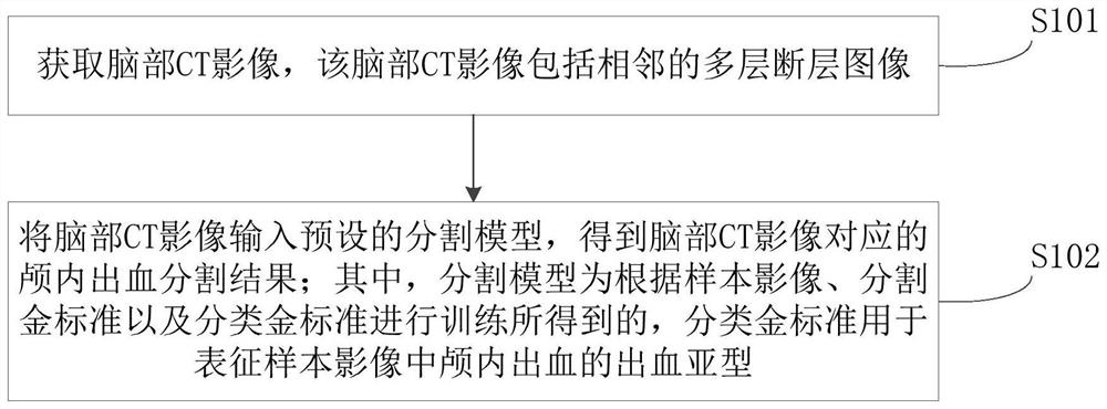
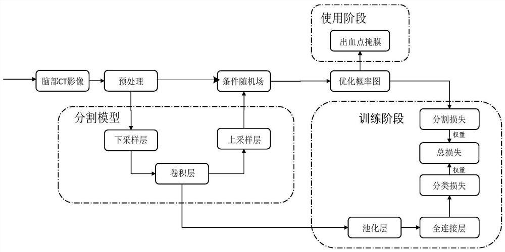
The invention relates to a brain image segmentation method and device, computer equipment and a readable storage medium. The method comprises the steps that a brain CT image is acquired, wherein the brain CT image comprises adjacent multi-layer sectional images; the brain CT image is input into a preset segmentation model to obtain an intracranial hemorrhage segmentation result corresponding to the brain CT image, wherein the segmentation model is obtained by training according to the sample image, a segmentation gold standard and a classification gold standard, and the classification gold standard is used for representing a bleeding subtype of intracranial bleeding in the sample image. According to the method, the classification loss is used for guiding the segmentation model to carry outtraining, so that the precision of the segmentation model obtained by training can be greatly improved, and the precision of a brain CT image segmentation result is further improved. Meanwhile, the brain CT image is an adjacent multi-layer sectional image, so that the context information among all layers of images can be fully considered, and the precision of the obtained brain CT image segmentation result is further improved.

Description

technical field [0001] The present application relates to the technical field of image processing, in particular to a brain image segmentation method, device, computer equipment and readable storage medium. Background technique [0002] Intracranial hemorrhage (ICH) refers to the hemorrhage caused by the rupture of blood vessels in the brain, which will cause damage to brain cells and at the same time compress the surrounding nerve tissue and cause other obstacles, which will have a greater impact on the patient's health. In the current field of clinical medicine, doctors usually determine the location of intracranial hemorrhage by watching the patient's CT images; with the continuous development of deep learning technology, there are already deep learning-based intracranial hemorrhage segmentation methods to assist doctors in determining the location of intracranial hemorrhage. To improve the doctor's work efficiency. [0003] Before using the deep learning network to segm...

Claims


Application Information

Patent Timeline
IPC IPC(8): G06T7/11G06K9/62G06N3/04G06N3/08
CPCG06T7/11G06N3/08G06T2207/10081G06T2207/30016G06N3/045G06F18/2415
Inventor 张朗廖术
Owner SHANGHAI UNITED IMAGING INTELLIGENT MEDICAL TECH CO LTD
Learn More