Method and system for identifying cells in embryo light microscope image, equipment and storage medium

A recognition method, a technology in the image, applied in the field of artificial intelligence, can solve the problems of no automatic and efficient detection and quantitative evaluation of the development of in vitro fertilized egg embryos, and the missed detection of high overlapping cells, so as to improve the accuracy and reduce the missing The effect of detection rate

Active Publication Date: 2020-12-11
SUN YAT SEN UNIV
View PDF7 Cites 5 Cited by
  • Summary
  • Abstract
  • Description
  • Claims
  • Application Information

AI Technical Summary

Problems solved by technology

NMS (non-maximum value suppression algorithm) is the basic structure of fasterrcnn, which is used to remove redundant overlapping detection frames in the model prediction stage. SoftNMS is an improvement to the original NMS algorithm, which performs better in the detection of low overlapping objects. However, it

Method used


Image

  • Method and system for identifying cells in embryo light microscope image, equipment and storage medium
  • Method and system for identifying cells in embryo light microscope image, equipment and storage medium
  • Method and system for identifying cells in embryo light microscope image, equipment and storage medium

Examples


no. 1 example

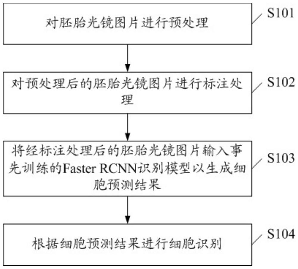
[0030] see figure 1 , figure 1 Shows the flow chart of the first embodiment of the cell identification method in the light microscope image of the embryo of the present invention, which includes:

[0031] S101, preprocessing the light microscope image of the embryo.

[0032] The light microscope pictures of embryos are taken by light microscope without dyeing treatment, so the light microscope pictures of embryos appear gray as a whole. Due to the transparency and serious overlap of the cells, the boundaries of the cells are blurred. At the same time, the brightness difference between the embryo light microscope images is small, and the foreground and background color distinction is insufficient, which has caused great difficulties for cell identification. .

[0033] In order to improve the foreground and background, as well as the color difference between each cell, the present invention uses a neighborhood histogram equalization method to preprocess the embryo light microsc...

no. 2 example

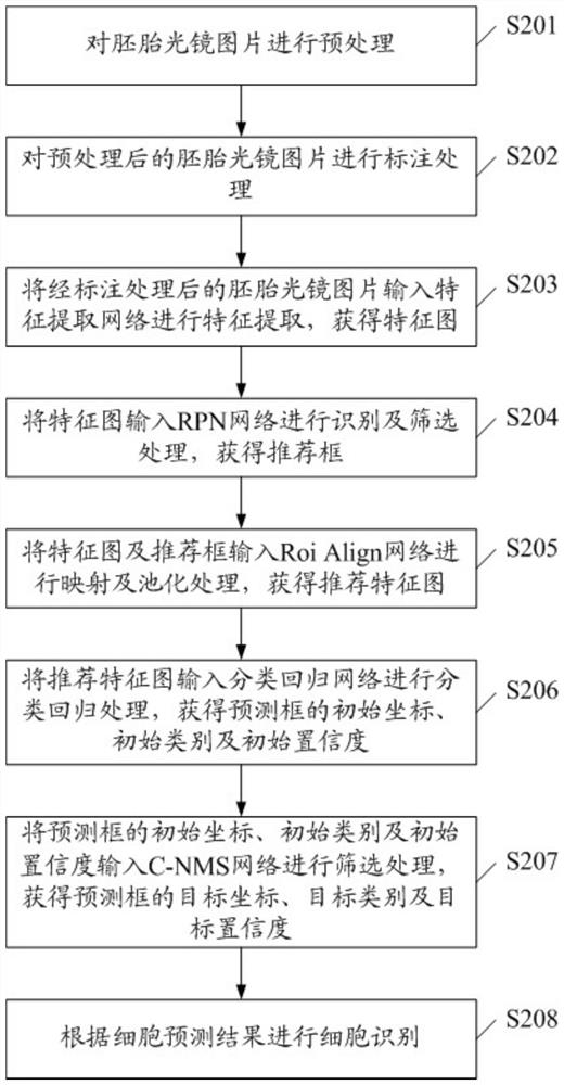
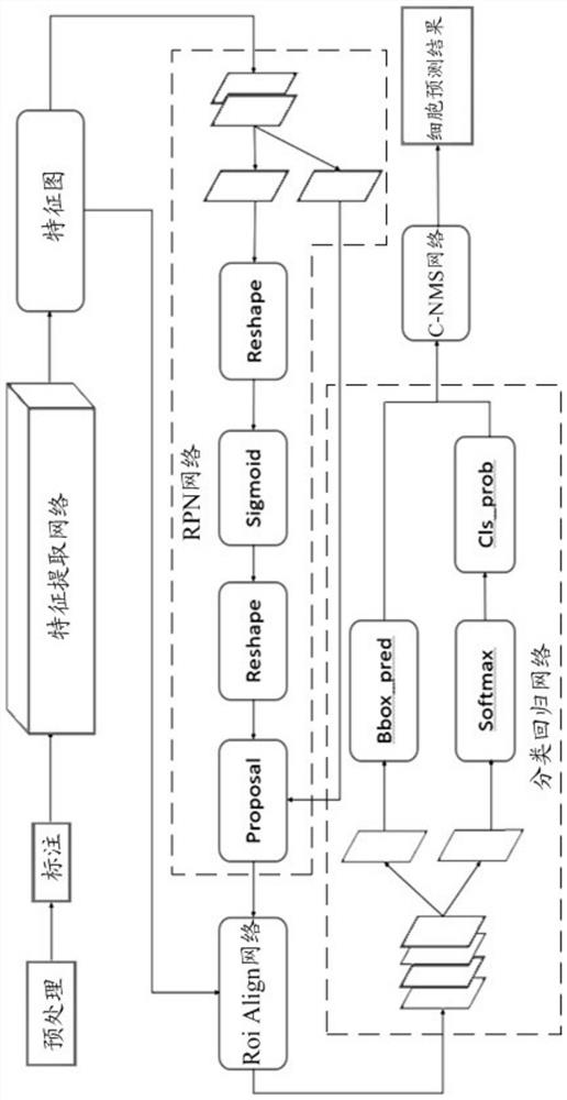
[0045] see figure 2 and image 3 , figure 2 and image 3 Shows the flow chart of the second embodiment of the cell identification method in the light microscope image of the embryo of the present invention, which includes:

[0046] S201, preprocessing the embryo light microscope image.

[0047] S202, labeling the preprocessed embryo light microscope image.

[0048] S203, inputting the marked embryo light microscope image into the feature extraction network for feature extraction to obtain a feature map.

[0049] In the prior art, the feature extraction network generally uses the VGG network. Different from the prior art, in the present invention, the feature extraction network is a ResNet50 full convolutional network. The ResNet50 network has deeper layers than the original VGG network, and has a residual structure, which is superior in feature extraction.

[0050] ResNet50 is a fully convolutional network with a total of 50 convolutional layers. The input of the Res...


PUM


Abstract

The invention discloses a method for identifying cells in an embryo light microscope image. The method comprises the following steps: preprocessing the embryo light microscope image; carrying out labeling processing on the preprocessed embryo light microscope picture; inputting the marked embryo light microscope picture into a FasterRCNN recognition model trained in advance to generate a cell prediction result, wherein the FasterRCNN recognition model comprises a feature extraction network, an RPN network, a Roi Align network, a classification regression network and a C-NMS network; and carrying out cell identification according to the cell prediction result. The invention further discloses a system for identifying the cells in the embryo light microscope image, computer equipment and a computer readable storage medium. By adopting the method and the system, accurate extraction of the cells in the embryo light microscope picture is realized through deep optimization of the Faster RCNNnetwork, meanwhile, a brand-new CNMS network is constructed, the detection score is flexibly adjusted through detection of the overlapping proportion and the area proportion of the detected objects, and the omission ratio is remarkably reduced.

Description

technical field [0001] The invention relates to the technical field of artificial intelligence, in particular to a cell recognition method in an embryo light microscope image, a cell recognition system in an embryo light microscope image, computer equipment and a computer storage medium. Background technique [0002] With the continuous advancement of medicine, the technology of test-tube baby and in vitro fertilization in the hospital is becoming more and more mature, and the number of cases of trying test-tube baby in the hospital is increasing, and the workload and intensity of reproductive doctors increase accordingly. Reproductive doctors perform quantitative detection and quality assessment of embryonic cells on embryonic light microscope images, which requires both highly accurate judgments and repeated image browsing. At present, the number detection and quality evaluation of embryonic cells are all done manually, and there is no corresponding automatic auxiliary tec...

Claims


Application Information

Patent Timeline
IPC IPC(8): G06K9/00G06K9/32G06N3/04G06N3/08
CPCG06N3/084G06V20/695G06V20/698G06V10/25G06N3/045
Inventor 王剑波李伟忠王文军张宁锋
Owner SUN YAT SEN UNIV
Why Eureka
Try Eureka