Cancer WSI segmentation method based on local classification neural network

A technology of neural network and classification network, which is applied in the field of segmentation of cancer full-view digital pathological slices, which can solve problems such as patient analysis, difficulty for doctors to pay attention to details, and large WSI size

Inactive Publication Date: 2020-12-18
FUDAN UNIV
View PDF9 Cites 6 Cited by
  • Summary
  • Abstract
  • Description
  • Claims
  • Application Information

AI Technical Summary

Problems solved by technology

Although professional doctors can complete the diagnosis by analyzing WSI, it is difficult for doctors to pay attention to all the details due to the huge size of WSI
At the same tim...

Method used


Image

  • Cancer WSI segmentation method based on local classification neural network
  • Cancer WSI segmentation method based on local classification neural network
  • Cancer WSI segmentation method based on local classification neural network

Examples


Embodiment Construction

[0025] The embodiments of the present invention will be described in detail below, but the protection scope of the present invention is not limited to the examples.

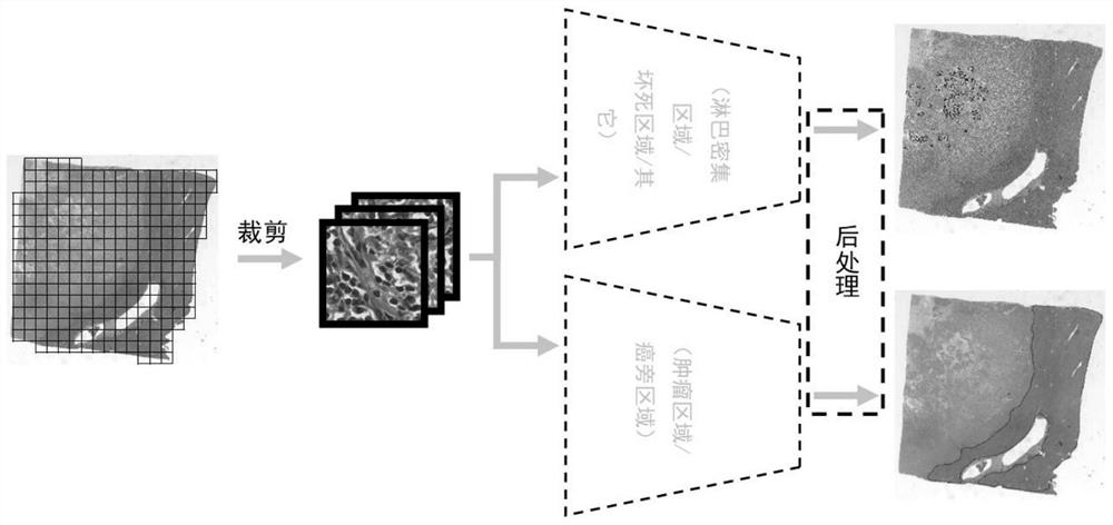
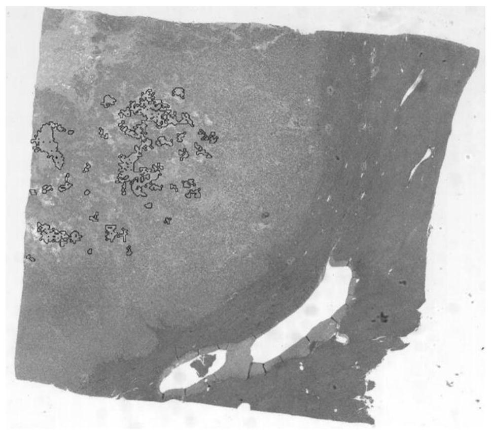
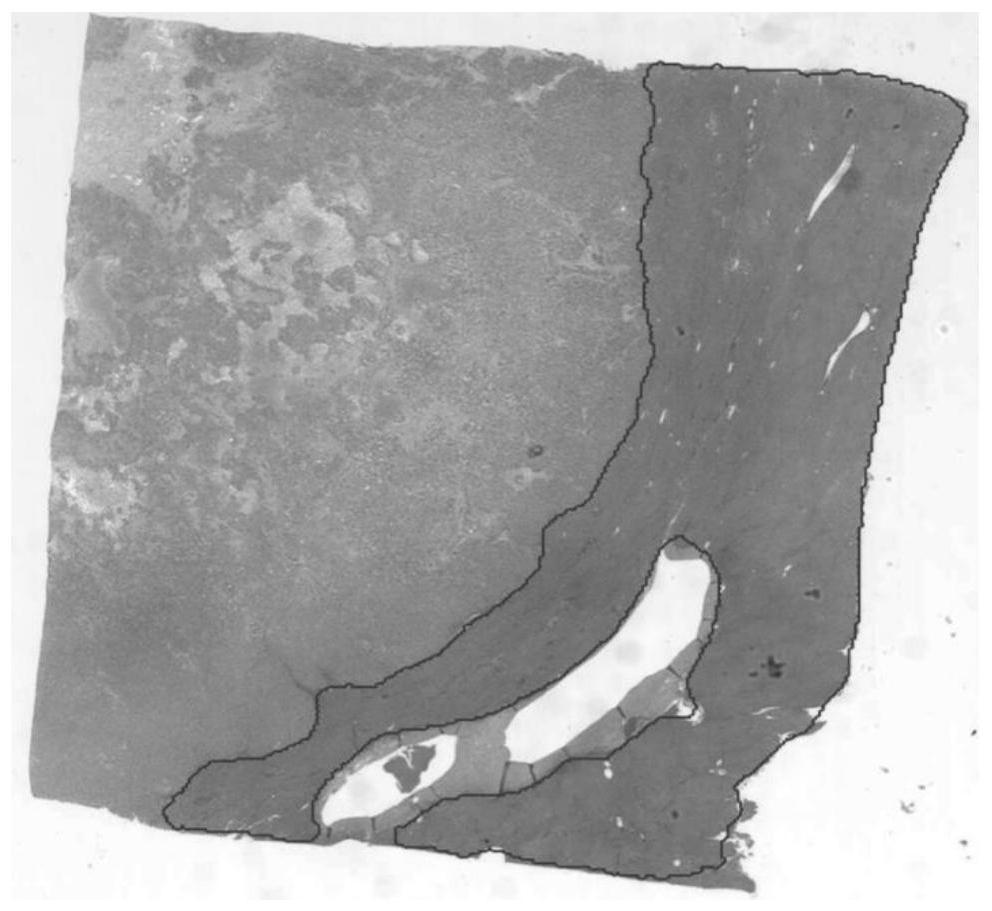
[0026] use figure 1 In the framework of the process, 86 pathological slices marked with lymphatic dense areas and necrotic areas, and 241 pathological slices marked with paracancerous and tumor areas were used to train two target detection neural networks to obtain automatic detection and diagnosis models.

[0027] The specific process is:

[0028] (1) Before training, the Otsu method was used to perform threshold segmentation on the green channel of the pathological slice to distinguish the background, so as to obtain the mask of the tissue area. Divide the WSI into several non-overlapping image blocks with a length and width of 256 pixels, and then randomly sample several image blocks from different regions in the mask as training samples according to manual annotation. The sampled image blocks require that ...


PUM


Abstract

The invention belongs to the technical field of medical image intelligent processing, and particularly relates to a cancer full-view digital pathological section segmentation method based on a local classification neural network, which comprises the following steps of: dividing a pathological section into a plurality of image blocks which have fixed sizes and are not overlapped; sending each imageblock into a classification model based on a convolutional neural network, judging the category of the block through forward propagation, and splicing the classification results of the blocks according to the positions of the blocks on the original image, thereby obtaining a heat map of different tissue distribution in the whole slice; finally, conducting median filtering on the heat map, removing an area with a too small area, so as to obtain a segmentation image and assist a doctor in diagnosis. By means of the method, the areas where different tissues in the whole pathological section arelocated can be rapidly marked, doctors are effectively assisted in diagnosis, and the diagnosis accuracy and efficiency are improved.

Description

technical field [0001] The invention belongs to the technical field of medical image intelligent processing, and in particular relates to a method for segmenting pathological slices, and more specifically, relates to a method for segmenting cancer full-field digital pathological slices based on a local classification neural network. Background technique [0002] With the development of whole slide scanning technology, a large number of tissue slices are scanned as whole-field digital pathological slides (Wholeslide image, WSI), stored in digital form, and widely used in cancer pathological diagnosis. Although professional doctors can complete the diagnosis by analyzing WSI, it is difficult for doctors to pay attention to all the details of WSI due to its huge size. At the same time, there are many factors that affect the prognosis of cancer, and it is difficult for doctors to analyze the relevant information of the patient's prognosis through pathological slides. [0003] B...

Claims


Application Information

Patent Timeline
IPC IPC(8): G06T7/11G06T7/136G06T5/20G06K9/62
CPCG06T7/11G06T7/136G06T5/20G06T2207/20081G06T2207/20084G06T2207/20032G06T2207/30096G06F18/24
Inventor 颜波高强谭伟敏丁光宇凌宇
Owner FUDAN UNIV
Why Eureka
Try Eureka