
Medical image fusion algorithm based on deep convolutional neural network

A neural network and deep convolution technology, applied in the field of medical image fusion, can solve the problems of not being able to provide the anatomical details of organs or lesions, not being able to reflect the function of organs, and poor resolution of functional images, so as to achieve good confidence and improve Effectiveness, the effect of increasing the success rate

Pending Publication Date: 2020-12-29
HARBIN UNIV OF SCI & TECH
View PDF0 Cites 2 Cited by
  • Summary
  • Abstract
  • Description
  • Claims
  • Application Information

AI Technical Summary

Problems solved by technology

They provide images of different modalities for medical diagnosis. These multimodal medical images can provide different medical information, each with its own advantages and disadvantages: the resolution of functional images is poor, but the organ function and metabolism information it provides is anatomical Irreplaceable by images; anatomical images provide high-resolution anatomical information of organs (functional images cannot provide anatomical details of organs or lesions), but cannot reflect the function of organs
Although there are many fusion methods of medical images at present, there are still many unsolved technical problems in clinical application.

Method used


Image

  • Medical image fusion algorithm based on deep convolutional neural network
  • Medical image fusion algorithm based on deep convolutional neural network
  • Medical image fusion algorithm based on deep convolutional neural network

Examples


Embodiment 1

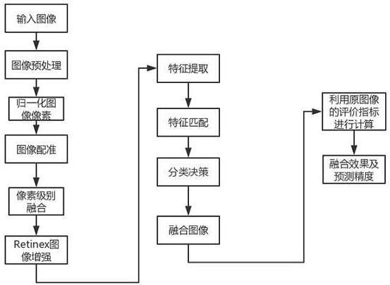
[0051] A medical image fusion algorithm based on a deep convolutional neural network, the algorithm includes the following steps:

[0052] Step 1. Input CT images and MRI images, and perform image preprocessing operations, including gray scale, binarization, filtering and denoising;

[0053] Step 2, normalize the two images, and integrate images from different sources at the same pixel level;

[0054] Step 3, using the best registration method to register the images, and using a search algorithm to find a solution that makes the similarity measure optimal in the search space;

[0055] Step 4. Perform pixel-level image fusion processing on the sub-band images after registration, and perform feature extraction and enhancement on the fused sub-band images through the multi-scale Retinex algorithm. Multi-scale Retinex is developed on the basis of the single-scale Retinex algorithm. , the specific process of the single-scale Retinex algorithm is as follows:

[0056] (1) Read in t...

Embodiment 2

[0081] According to the medical image fusion algorithm based on the deep convolutional neural network described in Embodiment 1, the step 3 performs pixel-level image fusion on the registered sub-band images using Contourlet transform.

[0082] The purpose of image fusion is to obtain a comprehensive image that conforms to human visual perception. At present, wavelet transform is widely used in the field of image fusion because of its good visual multi-scale and time-frequency local characteristics. However, for the two-dimensional image data structure, the wavelet transform cannot effectively represent the important details such as straight lines, curves, and edge directions. In multi-scale geometric analysis, the non-subsampled contourlet transform (NSCT) is called the true The sparsest representation of the image, it inherits the multi-scale and time-frequency locality of wavelet, and has multi-directionality and anisotropy, and solves the ringing effect caused by the subsam...


PUM


Abstract

The invention relates to a medical image fusion algorithm based on a deep convolutional neural network. At present, many medical image fusion methods exist, but many technical problems which are not solved yet exist in clinical application. Firstly, due to the fact that imaging principles of various imaging systems are different, image acquisition modes, formats, image sizes, image quality, imagespace and image time characteristics of the imaging systems are greatly different. The method comprises the following steps: inputting a CT image and an MRI image, and carrying out image preprocessingoperation; normalizing two images, and integrating the images of different sources under the same pixel level; registering images by adopting an optimal registration method, and finding a solution enabling similarity measurement to be optimal in a search space by using a search algorithm; and performing pixel-level image fusion on the registered sub-band images, and performing feature extractionenhancement on the fused sub-band images through a multi-scale Retinex algorithm. The invention is used for the medical image fusion algorithm based on the deep convolutional neural network.

Description

Technical field: [0001] The invention relates to the technical field of medical image fusion, in particular to a medical image fusion algorithm based on a deep convolutional neural network. Background technique: [0002] With the development of medicine and computer technology, medical imaging has become a routine auxiliary means for clinicians to diagnose, treat or formulate surgical plans, whether it is disease diagnosis, determination and simulation of surgical plans, or the implementation and monitoring of surgical plans. It is inseparable from the accurate information given by medical images. Different medical images can provide different information on related organs and tissues of the human body, and the morphological and functional information obtained by different imaging technologies on the same anatomical structure of the human body are often mutually different and complementary. Therefore, using image fusion technology, different medical imaging information can ...

Claims


Application Information

Patent Timeline
Patent Type & Authority Applications(China)
IPC IPC(8): G06T7/90G06T5/50G06N3/04G06K9/62G16H30/20
CPCG06T5/50G06T7/90G16H30/20G06T2207/20221G06N3/045G06F18/214G06F18/251
Inventor 何召兰姚徐丁淑培
Owner HARBIN UNIV OF SCI & TECH
Learn More