
Multi-mode automatic ventricular segmentation system and using method thereof

A multi-modal, ventricular technology, applied in the field of bioinformatics, can solve problems such as inaccurate automatic segmentation, and achieve the effects of reducing manpower consumption, low labeling costs, and easy access

Pending Publication Date: 2021-01-08
THE SECOND PEOPLES HOSPITAL OF SHENZHEN
View PDF0 Cites 4 Cited by
  • Summary
  • Abstract
  • Description
  • Claims
  • Application Information

AI Technical Summary

Problems solved by technology

[0005] The purpose of the present invention is to provide a multimodal automatic ventricle segmentation system and its use method to solve the problem of inaccurate automatic segmentation of existing brain medical imaging images

Method used


Image

  • Multi-mode automatic ventricular segmentation system and using method thereof
  • Multi-mode automatic ventricular segmentation system and using method thereof
  • Multi-mode automatic ventricular segmentation system and using method thereof

Examples


Embodiment Construction

[0051] The multimodal automatic ventricle segmentation system proposed by the present invention and its usage method will be further described in detail below in conjunction with the accompanying drawings and specific embodiments. Advantages and features of the present invention will be apparent from the following description and claims. It should be noted that all the drawings are in a very simplified form and use imprecise scales, and are only used to facilitate and clearly assist the purpose of illustrating the embodiments of the present invention.

[0052] In addition, unless otherwise stated, features in different embodiments of the present invention can be combined with each other. For example, a feature in the second embodiment may be used to replace a corresponding or functionally identical or similar feature in the first embodiment, and the resulting embodiment also falls within the scope of disclosure or description of the present application.

[0053] The core idea...


PUM


Abstract

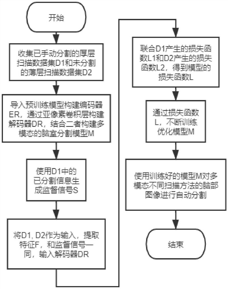
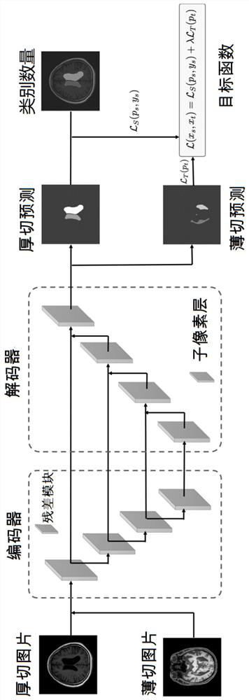
The invention provides a multi-mode automatic ventricular segmentation system and a use method thereof. The method comprises the following steps of collecting a manually segmented thick layer scanningdata set D1 and an unsegmented thin layer scanning data set D2; importing a pre-training model to construct an encoder ER, constructing a decoder DR through a sub-pixel convolution layer, and constructing a multi-modal ventricular segmentation model M in combination with the encoder ER and the decoder DR; generating a supervision signal S by using segmented information in the thick-layer scanningdata set D1; taking the thick-layer scanning data set D1 and the thin-layer scanning data set D2 as input, extracting a feature F, and inputting the feature F and a supervision signal S into a decoder DR; combining a loss function L1 generated by the thick-layer scanning data set D1 and a loss function L2 generated by the thin-layer scanning data set D2 to obtain a loss function L of the ventricular segmentation model M; continuously training and optimizing the ventricular segmentation model M according to the loss function L; and using the trained ventricular segmentation model M to automatically segment the brain images of different multi-modal scanning methods.

Description

technical field [0001] The invention relates to the technical field of biological information, in particular to a multimodal automatic ventricle segmentation system and a method for using the same. Background technique [0002] Ventricular volume is closely related to many brain diseases, and many studies have proposed that altered ventricular volume is a feature of diseases such as schizophrenia, Parkinson's disease, Alzheimer's disease, hydrocephalus and brain atrophy. Accurate measurement of ventricular volume has very important clinical significance in detecting early diseases, evaluating patients' conditions, diagnosing diseases, and evaluating surgical effects. Therefore, measuring the volume of the ventricle has very important value in the medical field. [0003] Ventricular segmentation is the only way to measure ventricular volume. It refers to the use of appropriate methods to divide the ventricles of the scanned medical imaging images of the patient's brain. It...

Claims


Application Information

Patent Timeline
IPC IPC(8): G06T7/10G06N3/04G06K9/62
CPCG06T7/10G06T2207/30016G06T2207/10081G06T2207/10088G06T2207/20081G06T2207/20084G06N3/045G06F18/214
Inventor 夏军杨光牛张明江荧辉叶晴昊王旻皓
Owner THE SECOND PEOPLES HOSPITAL OF SHENZHEN
Learn More