

IV-OCT blood vessel contour recognition method based on uniqueness of tube wall connected domain
A technology of connected domains and blood vessels, applied in the field of fully automatic morphological recognition of lumen contours, can solve problems such as poor shooting conditions and difficulty in extracting lumen contours, and achieve low signal-to-noise ratio effects
- Summary
- Abstract
- Description
- Claims
- Application Information
AI Technical Summary
Problems solved by technology
Method used
Image

Examples
Embodiment Construction
[0059] The present invention is based on the recognition method proposed by reference 14, and its steps are simplified to a certain extent, some steps are inherited and improved, and a new processing method is proposed. The present invention inherits the basic image processing methods such as the morphological opening operation used by it; abandons the algorithm of dynamic segmentation; improves the method of morphological direction and area detection and the method of signal retrieval by secondary recognition, and is developed in Dajin Based on the binarization of the method, the processing of partitioning and judging the existence of the vessel wall is proposed; in addition, a new method of removing residual blood and catheters based on the uniqueness of the connected domain of the vessel wall on the A-line is proposed, which effectively improves The recognition rate and the robustness of the method are improved.
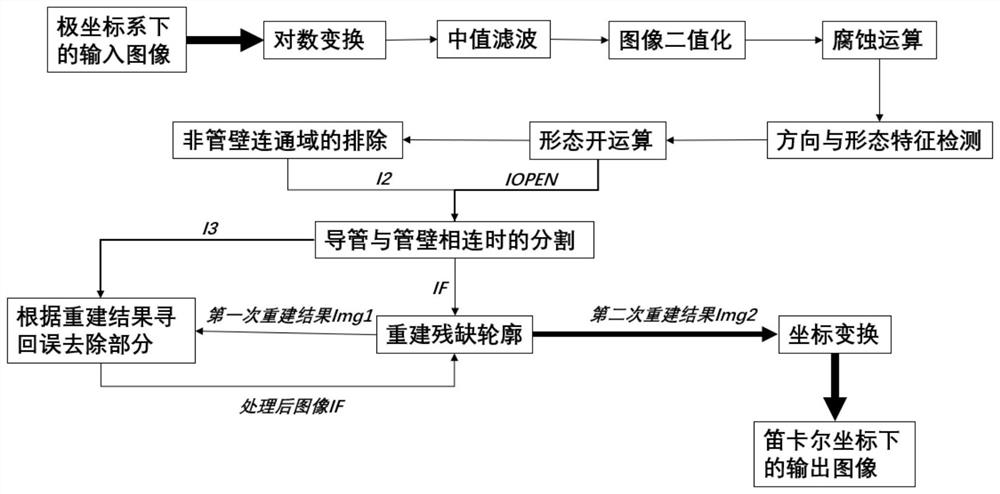
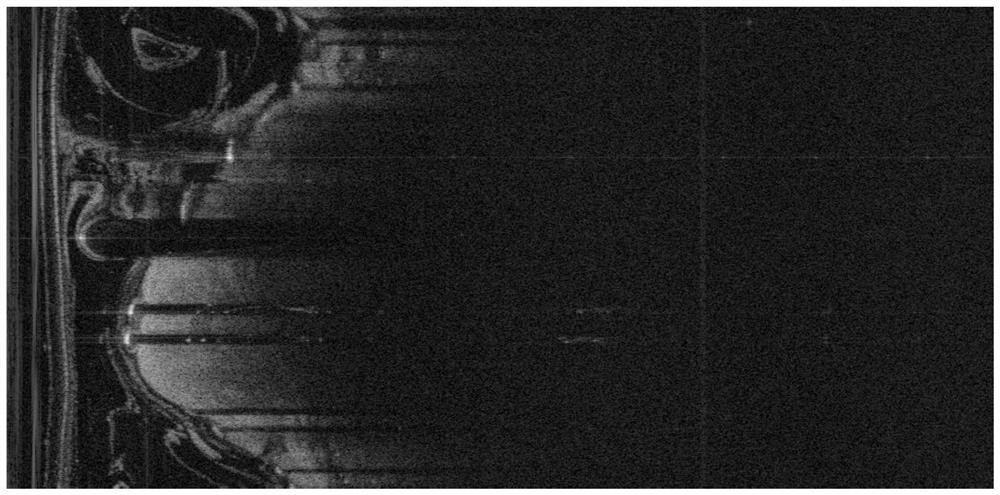
[0060] Such as figure 1 As shown, the lumen outline automat...
PUM
 Abstract
Description
Claims
Application Information