Eureka AIR delivers breakthrough ideas for toughest innovation challenges, trusted by R&D personnel around the world.

Whole organ cell proliferation quantitative detection method

A quantitative detection method and cell proliferation technology, applied in instruments, measuring devices, scientific instruments, etc., can solve problems such as difficult overall cell proliferation

Pending Publication Date: 2021-02-05
RES CENT FOR ECO ENVIRONMENTAL SCI THE CHINESE ACAD OF SCI
View PDF4 Cites 0 Cited by
  • Summary
  • Abstract
  • Description
  • Claims
  • Application Information

AI Technical Summary

Problems solved by technology

[0004] In view of the above problems, the present invention provides a method for quantitative detection of cell proliferation in whole organs, which is used to at least partially solve technical problems such as the difficulty in quantitatively analyzing the overall cell proliferation of tissues by traditional methods

Method used


Image

  • Whole organ cell proliferation quantitative detection method
  • Whole organ cell proliferation quantitative detection method
  • Whole organ cell proliferation quantitative detection method

Examples


Embodiment Construction

[0022] In order to make the object, technical solution and advantages of the present invention clearer, the present invention will be described in further detail below in conjunction with specific embodiments and with reference to the accompanying drawings.

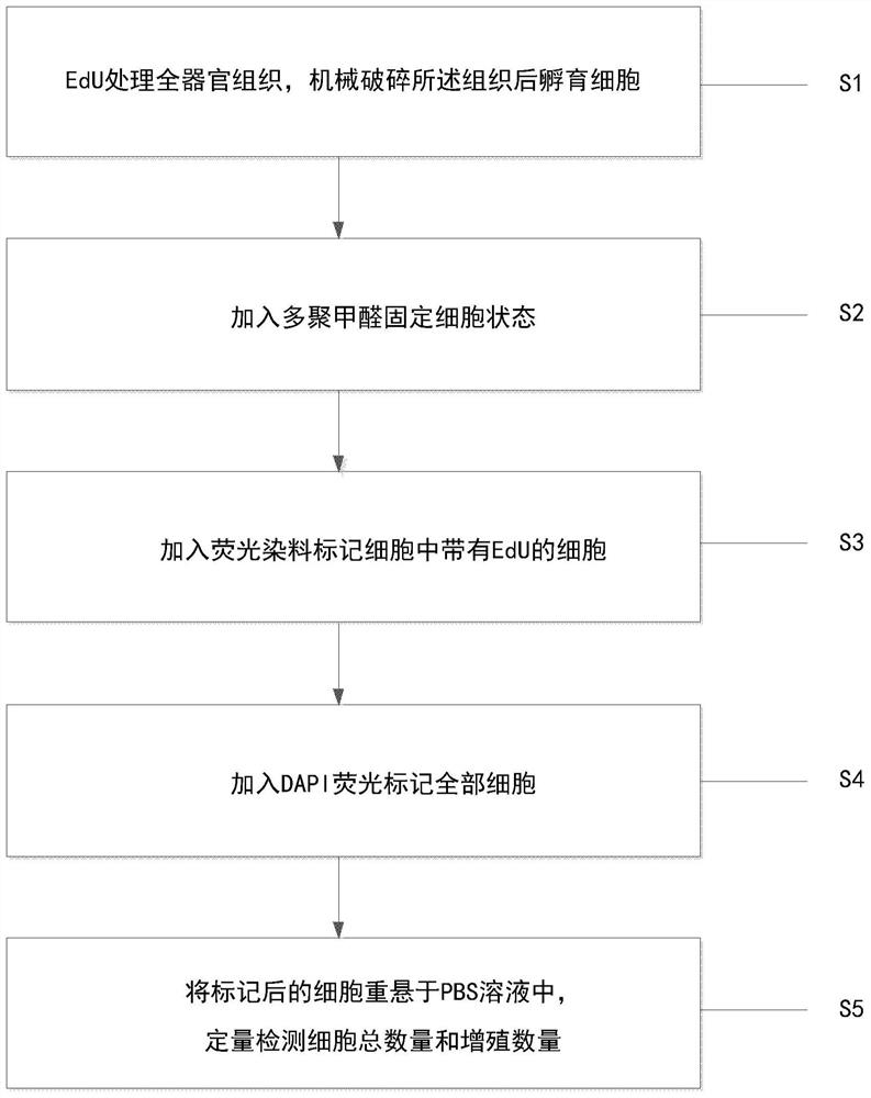
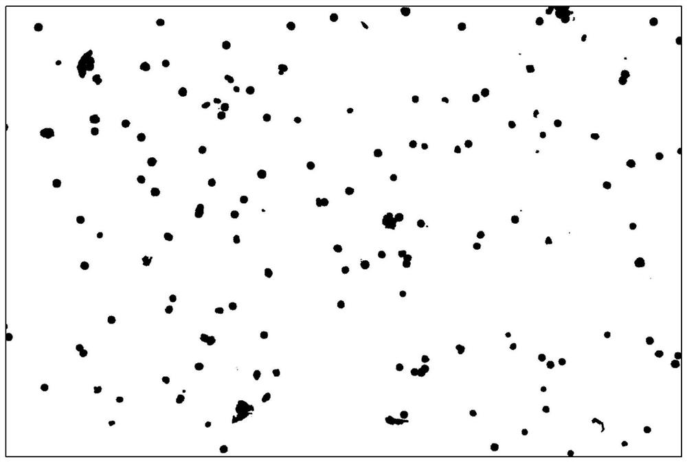
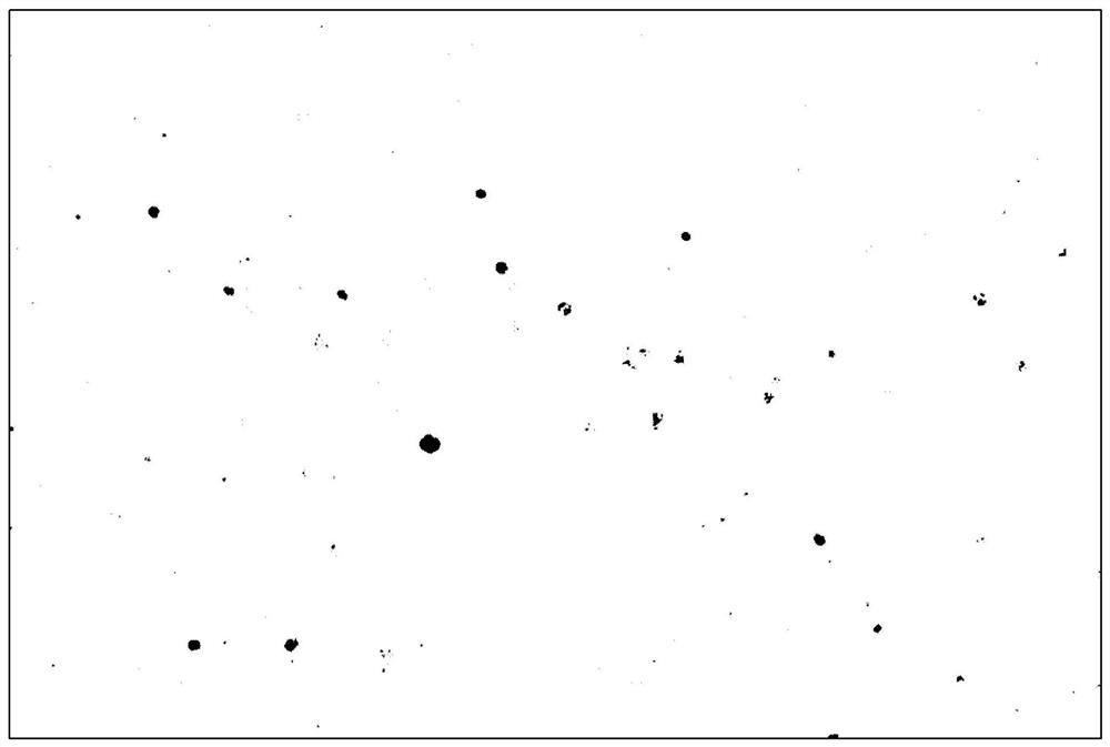
[0023] The embodiment of the present invention provides a method for quantitative detection of whole organ cell proliferation, please refer to figure 1 , including: S1, EdU treatment of whole organ tissue, mechanically breaking the tissue and incubating the cells; S2, adding paraformaldehyde to fix the cell state; S3, adding fluorescent dye to label the cells with EdU in the cells; S4, adding DAPI fluorescent labeling to all the cells ; S5, the labeled cells were resuspended in PBS solution, and the total number of cells and the number of proliferation were quantitatively detected.

[0024] EdU can be inserted into the DNA molecule being replicated during cell proliferation. Based on the conjugation reaction of EdU and dy...


PUM

PropertyMeasurementUnit
Concentrationaaaaaaaaaa

Abstract

The invention provides a whole organ cell proliferation quantitative detection method, which comprises the following steps of: S1, treating whole organ tissues by means of EdU, mechanically crushing the tissues, and incubating the cells; S2, adding paraformaldehyde into the cells to fix a cell state; S3, adding a fluorescent dye to mark cells with EdU in the cells; S4, adding DAPI fluorescence tomark all cells; and S5, re-suspending the marked cells in a PBS solution, and quantitatively detecting the total number and proliferation number of the cells. The whole organ cell proliferation quantitative detection method provided by the invention avoids the problem that the traditional slice detection method is difficult to quantitatively analyze the overall tissue cell proliferation, and is very suitable for quantitatively analyzing the cell proliferation condition of the heterogeneous tissue.

Description

technical field [0001] The invention relates to the technical field of cell detection, in particular to a quantitative detection method for whole organ cell proliferation. Background technique [0002] For traditional cell proliferation detection techniques, frozen section or paraffin section is usually used to obtain a section of the target tissue, and then the cell proliferation of the whole tissue is analyzed by marking the proliferating cells on the section. For heterogeneous tissues such as brain matter, the method of tissue sectioning often results in inconsistent proportions of proliferating cells in different brain sections, which affects qualitative analysis. In addition, due to the difficulty of obtaining a relatively consistent section of brain tissue, it is difficult to quantitatively analyze the overall cell proliferation of the tissue. Contents of the invention [0003] (1) Technical problems to be solved [0004] In view of the above problems, the present ...

Claims


Application Information

Patent Timeline
IPC IPC(8): G01N15/14G01N1/28G01N1/30
CPCG01N15/1434G01N1/286G01N1/30G01N2015/1486G01N2015/144
Inventor 秦占芬董梦琦
Owner RES CENT FOR ECO ENVIRONMENTAL SCI THE CHINESE ACAD OF SCI
Why Eureka
Eureka Blog
Learn More