Supercharge Your Innovation With Domain-Expert AI Agents!

Endoscopic imaging method, device, equipment and storage medium

An imaging method and technology of imaging equipment, applied in the field of endoscopy, can solve the problems of low contrast in the lesion area, inability to obtain inspection results, and easy missed diagnosis.

Active Publication Date: 2021-05-14
SONOSCAPE MEDICAL CORP
View PDF5 Cites 0 Cited by
  • Summary
  • Abstract
  • Description
  • Claims
  • Application Information

AI Technical Summary

Problems solved by technology

[0004] However, when endoscopic imaging is performed under white light illumination mode, the contrast between the lesion area and the normal mucosa is low, and it is easy to miss a diagnosis. Moreover, once a suspected lesion is missed under white light illumination mode, it will not be switched to special light illumination in the future. Extensive inspection of the model will eventually lead to the inability to obtain more accurate inspection results

Method used


Image

  • Endoscopic imaging method, device, equipment and storage medium
  • Endoscopic imaging method, device, equipment and storage medium
  • Endoscopic imaging method, device, equipment and storage medium

Examples


Embodiment Construction

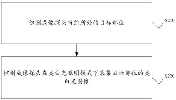
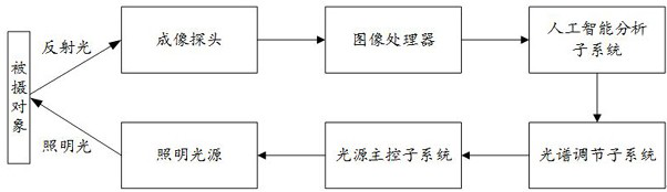
[0061] The core of the present application is to provide an endoscopic imaging method, which can be applied to endoscopic imaging equipment, and the endoscopic imaging equipment includes an imaging probe that can be placed in the body. This method can be applied to any endoscopic examination scene, such as endoscopic examination of the digestive tract. For the convenience of description, the embodiment of the present application mainly uses the endoscopic examination of the digestive tract as an example for illustration.

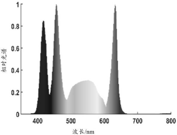
[0062] In related technologies, when endoscopic imaging is performed under white light illumination mode, the contrast between the lesion area and the normal mucosa is low, and misdiagnosis is likely to occur. In the special lighting mode, a fixed spectral lighting imaging is used, which does not take into account the differences in tissue optical characteristics of various anatomical parts of the digestive tract, resulting in large differences in the contra...


PUM


Abstract

This application discloses an endoscopic imaging method. The method includes the following steps: identifying the target part where the imaging probe is currently located; controlling the imaging probe to collect a white-like light image of the target part in a white-like light illumination mode, in which the color tone of the white-like light image is The tone of the white-light image of the target part is similar. In the same image area, the contrast between the background information and the detailed information in the white-light image in the blue channel is greater than the contrast between the background information and the detailed information in the white-light image in the blue channel. Applying the technical solution provided by this application, when performing endoscopic imaging, it can present an imaging tone similar to a white light image, which is convenient for users to identify. At the same time, it can effectively enhance the contrast between the lesion area and normal mucosa in each part, and reduce the missed detection rate. , improve the accuracy of inspection results. This application also discloses an endoscopic imaging device, equipment and computer storage medium, which have corresponding technical effects.

Description

technical field [0001] The present application relates to the technical field of endoscopes, in particular to an endoscope imaging method, device, equipment and storage medium. Background technique [0002] With the advancement of medical technology, various inspection methods are gradually increasing, and the inspection results provide an important basis for subsequent diagnosis, and the accuracy of the inspection results is even more important. [0003] In endoscopic examination, because the white light illumination mode is closer to the visual effect of the human eye, doctors usually use the white light illumination mode to conduct a full inspection of the digestive tract. After suspicious lesions are found, they switch to NBI (Narrow Band Imaging, Narrow Band Imaging), BLI (Blue Laser Imaging, blue laser imaging), LCI (Linked Color Imaging, linkage imaging) and other special lighting modes for further detailed examination. [0004] However, when endoscopic imaging is pe...

Claims


Application Information

Patent Timeline
Patent Type & Authority Patents(China)
IPC IPC(8): A61B1/06A61B1/04A61B1/00H04N5/225H04N5/232H04N5/235
CPCA61B1/0638A61B1/0684A61B1/00009A61B1/04H04N23/555H04N23/56H04N23/80H04N23/74
Inventor 汪洋邱建军王森豪陈云亮张雅馨
Owner SONOSCAPE MEDICAL CORP
Features
  • R&D
  • Intellectual Property
  • Life Sciences
  • Materials
  • Tech Scout
  • Unparalleled Data Quality
  • Higher Quality Content
  • 60% Fewer Hallucinations
Learn More