Multichannel retinal vessel image segmentation method based on U-net network

A retinal blood vessel and image segmentation technology, applied in image analysis, image enhancement, image data processing, etc., can solve problems such as inability to make good use of context information, incomplete feature extraction, small receptive field of convolution operation, etc., and achieve relief Insufficient image segmentation and mis-segmentation problems, beneficial feature learning, high segmentation sensitivity and accuracy

Pending Publication Date: 2021-03-09
HANGZHOU DIANZI UNIV
View PDF0 Cites 4 Cited by
  • Summary
  • Abstract
  • Description
  • Claims
  • Application Information

AI Technical Summary

Problems solved by technology

However, the problems existing in the traditional FCN are also obvious: its network structure classifies pixels, so the connection between pixels is inevitably ignored, and the receptive field of the convolution operation is too small to make good use of the context information, resulting in Incomplete feature extraction

Method used


Image

  • Multichannel retinal vessel image segmentation method based on U-net network
  • Multichannel retinal vessel image segmentation method based on U-net network
  • Multichannel retinal vessel image segmentation method based on U-net network

Examples


Embodiment Construction

[0056] The present invention will be further described below in conjunction with specific embodiments. The following description is only for illustration and acceptance, and does not limit the present invention in any form.

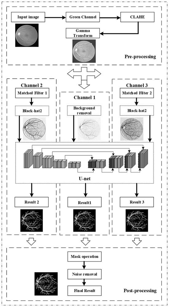
[0057] Such as figure 1 As shown, the implementation steps of the embodiments of the present invention are as follows:

[0058] Step 1. Perform data amplification operations on the training sets of the existing public datasets DRIVE, STARE, and CHASE_DB1. Specifically, perform horizontal flips, vertical flips, and 180-degree rotations on the images to increase the amount of data to 4 times the original. Among them, the STARE dataset randomly selects 15 images as the training set, and the CHASE_DB1 dataset selects the first 20 images as the training set.

[0059] Step 2, the preprocessing process of the image includes:

[0060] Step 2-1: Perform channel separation on the color image, select the green channel with better blood vessel clarity as the input i...


PUM


Abstract

The invention discloses a multichannel retinal vessel image segmentation method based on a Unet network. The method comprises the following steps: firstly, performing amplification processing and a series of preprocessing on a data set image to improve the image quality; secondly, combining a multi-scale matched filtering algorithm with an improved morphological algorithm to construct a multi-channel feature extraction structure of the Unet network; and then, carrying out network training on the three channels to obtain a required segmentation network, and carrying out adaptive threshold processing on an output result. According to the method, the Unet network and the multi-scale matched filtering algorithm are combined, compared with a pure Unet network, more blood vessel features can beextracted, higher segmentation accuracy and sensitivity are achieved, and the problems of insufficient segmentation and wrong segmentation of small blood vessels of the retinal blood vessel image arerelieved.

Description

technical field [0001] The invention relates to an image segmentation method, in particular to an image segmentation method improved by combining a U-net network in deep learning with an image matching filtering method. This method is practically applied in the segmentation of retinal blood vessel images. Background technique [0002] At present, the algorithms of image segmentation can be mainly divided into two categories: supervised learning methods and unsupervised learning methods. The unsupervised learning method mainly uses some characteristics of the image itself, manually sets the process and method of feature extraction, and has good segmentation results for the unique attributes of some images, but the use of this method largely depends on previous experience. , does not have good generalization when facing different images for segmentation processing. The most important feature of the supervised learning method is that the artificial segmentation results of the...

Claims


Application Information

Patent Timeline
Patent Type & Authority Applications(China)
IPC IPC(8): G06T7/11G06T7/155G06T5/00G06T5/40G06K9/46G06K9/62G06N3/04G06N3/08
CPCG06T7/11G06T7/155G06T5/002G06T5/40G06N3/088G06T2207/20024G06T2207/20221G06T2207/30041G06T2207/30101G06V10/443G06V10/462G06N3/045G06F18/214
Inventor 马玉良祝真滨李雪席旭刚张卫
Owner HANGZHOU DIANZI UNIV
Why Eureka
Try Eureka