Image attenuation correction method and application thereof

An attenuation correction, image technology, applied in image enhancement, image analysis, image data processing and other directions, can solve problems such as unclear images, increase the receptive field, and improve the effect of extraction

Pending Publication Date: 2021-03-16
NAT INST OF ADVANCED MEDICAL DEVICES SHENZHEN
View PDF0 Cites 0 Cited by
  • Summary
  • Abstract
  • Description
  • Claims
  • Application Information

AI Technical Summary

Problems solved by technology

[0006] Based on the problem that the image obtained by using computer tomography to provide anatomical information for attenuation correction method is not clear, this application provides an image attenuation correction method and its application

Method used


Image

  • Image attenuation correction method and application thereof
  • Image attenuation correction method and application thereof
  • Image attenuation correction method and application thereof

Examples


Embodiment

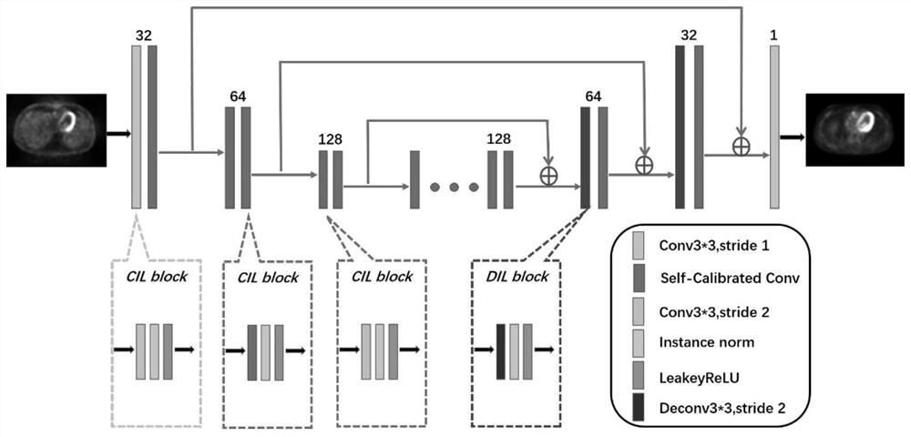
[0054] A self-correcting convolutional neural network for PET image attenuation correction, the specific operation steps are as follows:

[0055] Step 1: The generator network structure designed by this PET image attenuation correction network

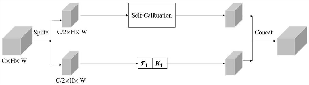
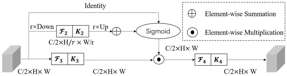
[0056] This module is a codec network with skip connections: all codecs are composed of a series of 2D convolutions. Adding skip connections is to speed up the process of network training and preserve more details of the image. The generator network contains a total of 17 layers, including 2 layers of convolution, 2 layers of deconvolution and 13 layers of self-correcting convolution. Each layer contains convolution layer / self-correction convolution / deconvolution layer, batch normalization layer and activation function layer. All convolution kernels used are of size 3×3. And the number of convolution kernels is 32, 32, 64, 64, 128, 128, 128, 128, 128, 128, 128, 128, 64, 64, 32, 32, 1 in sequence. And the convolution step size is 1....


PUM


Abstract

The invention belongs to the technical field of image imaging, and particularly relates to an image attenuation correction method and application thereof. At present, an image obtained by a method forproviding anatomical information for attenuation correction by computer tomography is not clear. For the synthesis from an NAC-PET image to an AC-PET image, self-correcting convolution can improve the extraction of the NAC-PET image content information by a generator and increase receptive field of convolutional layer (the receptive field being size of region mapped by pixel points on feature mapoutput by each layer of convolutional neural network (CNN) on original input image); and after the receptive field is increased, the pixel points on the feature map subjected to convolution extraction can better reflect information in the original image, and attenuation correction can be better carried out on a non-attenuation correction image (NAC-PET).

Description

technical field [0001] The present application belongs to the field of image imaging technology, and in particular relates to an image attenuation correction method and its application. Background technique [0002] Positron emission tomography (PET) plays an indispensable role in early detection of diseases and postoperative staging diagnosis, and is a functional imaging method widely used in neuroscience research. Increased accumulation of fluoro-D-glucose (FDG) used in PET relative to normal tissue is a useful marker of many cancers, helping to detect and localize malignancies. Despite its many advantages, PET imaging has several disadvantages that make it difficult to treat. Radioactive components may pose a risk to pregnant or nursing patients. Also, PET is a relatively new medical procedure that can be expensive. As such, it is still not offered in most medical centers around the world. Difficulties in providing PET imaging as part of treatment have increased the n...

Claims


Application Information

Patent Timeline
Patent Type & Authority Applications(China)
IPC IPC(8): G06T11/00G06T5/00G06T5/50G06N3/04G06N3/08
CPCG06T11/005G06T5/002G06T5/50G06N3/08G06T2207/10081G06T2207/10104G06T2207/20221G06T2207/20081G06T2207/20084G06T2207/30096G06N3/045
Inventor 郑海荣江洪伟李彦明万丽雯薛恒志胡战利
Owner NAT INST OF ADVANCED MEDICAL DEVICES SHENZHEN
Why Eureka
Try Eureka