A method to determine a degree of abnormality, a respective computer readable medium and a distributed cancer analysis system

A level and abnormal technology, applied in the field of distributed cancer analysis system, can solve the problems of damage robustness and classification, and achieve the effect of cost reduction and fast processing speed

Pending Publication Date: 2021-03-23
H LABS股份有限公司
View PDF0 Cites 1 Cited by
  • Summary
  • Abstract
  • Description
  • Claims
  • Application Information

AI Technical Summary

Problems solved by technology

[0015] Finally, adding new knowledge to neural networks is a big problem because robustness and classification can be destroyed by overfitting the training data

Method used


Image

  • A method to determine a degree of abnormality, a respective computer readable medium and a distributed cancer analysis system
  • A method to determine a degree of abnormality, a respective computer readable medium and a distributed cancer analysis system
  • A method to determine a degree of abnormality, a respective computer readable medium and a distributed cancer analysis system

Examples


Embodiment Construction

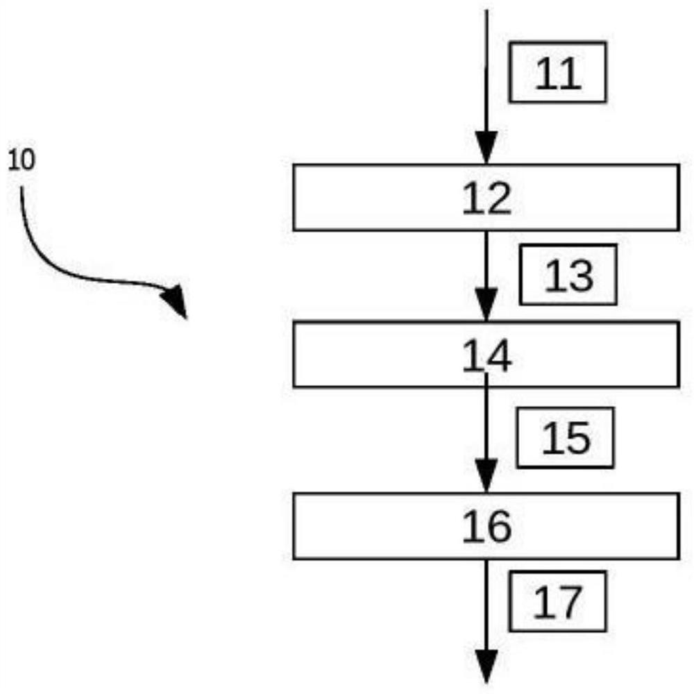
[0115] figure 1 A flow chart of a method of determining the degree of abnormality value 10 is shown. In a first step, the whole slice image 11 is processed during a segmentation stage 12 . Whole slide image 11 depicts a portion of human cells that may be cancerous. Furthermore, the whole slice image 11 may show cells that have been treated with a biomarker (eg CINTEC detection). As a result, discoloration appears in certain areas in the whole slide image, indicating certain chemical reactions. The full slice image 11 is then segmented into a plurality of image segments 13 in a segmentation stage 12 . Each image patch represents a portion of a full-slice image. Thus, a plurality of image segments 13 together form a full slice image 11 . Preferably, the image blocks 13 have the same size. In this embodiment, the size of the image blocks 13 is 30×30 pixels. In other embodiments, other block sizes are possible, such as dimensions of 100x100 pixels, 200x200 pixels, or 1000x1...


PUM


Abstract

Current cancer screening methods are not suitable to be applied on a broad scale and are not transparent to the patient. The problem is solved by a method to determine a degree of abnormality, the method comprising the following steps: a) receiving a whole slide image (11, w, 722), the whole slide image (11, w, 733) depicting at least a portion of a cell; b) classifying at least one image tile (13, 601, 721, 721', 721'') of the whole slide image (11, w, 722) using a neural network (600) to determine a local abnormality degree value (15, a_j, 519, 719, 719', 719'') associated with the at leastone image tile (13, 601, 721, 721', 721''), the local abnormality degree value (15, a_j, 519, 719, 719', 719'') indicating a likelihood that the associated at least one segment depicts at least a partof a cancerous cell; and c) determining a degree of abnormality (17) for the whole slide image (11, w, 722) based on the local abnormality degree value (15, a_j, 519, 719, 719', 719'') for the at least one image tile (13, 601, 721, 721', 721'').

Description

technical field [0001] The present application relates to a method of determining the degree of abnormality, a corresponding computer readable medium and a distributed cancer analysis system. Background technique [0002] Cancer screening programs rely on consistent early detection of cancer lesions by trusted experts. If the cancer is detected early enough, it can be treated locally and thus often effectively avoids the risk to the patient's health. In many cases, cancer screening involves taking a biopsy, which is a small sample of tissue from a potentially cancerous area. This biopsy is usually done during a routine medical checkup, or as specifically indicated after a prior medical checkup. As with any other pathological tissue specimen, biopsies are evaluated after preparation on glass slides by an expert (usually a board-certified pathologist). Pathologists are partly trained by a body of available specialists and are solely responsible for diagnosis. Often, pathol...

Claims


Application Information

Patent Timeline
Patent Type & Authority Applications(China)
IPC IPC(8): G06K9/00G06K9/46G06K9/62H04L9/32G06N3/04G06N3/08G06N5/02G06V10/764
CPCG06N3/08G06N5/02G06T7/0012G06T2207/10056G06T2207/10024G06T2207/20021G06T2207/20084G06T2207/20081G06T2207/30024G06T2207/30096H04L2209/88H04L9/3239G16H50/20G16H30/40G16H10/40G06F21/00G06T7/00G16H10/00H04L9/3236G06V20/698G06V10/454G06V10/82H04L9/50G06V10/764G06N3/042G06N3/045G16H30/20G06N3/04G06V2201/03G06F18/241G06F18/2178G06F18/2193
Inventor 贝尔德·拉尔曼
Owner H LABS股份有限公司
Why Eureka
Try Eureka