
Retina and optic nerve protective electrical stimulation device

A technique of retinal and electrical stimulation, applied in the direction of electrodes, electrotherapy, artificial respiration, etc.

Pending Publication Date: 2021-03-30
HARBIN MEDICAL UNIVERSITY
View PDF0 Cites 0 Cited by
  • Summary
  • Abstract
  • Description
  • Claims
  • Application Information

AI Technical Summary

Problems solved by technology

[0006] The present invention provides a retinal and optic nerve protective electrical stimulation device to solve the existing problems in the prior art presented in the above-mentioned background technology. Research on non-invasive eye transretinal electrical stimulation or transorbital electrical stimulation and its electrical stimulation device is of great importance to ophthalmology. Issues of great significance to the development of medicine

Method used


Image

  • Retina and optic nerve protective electrical stimulation device
  • Retina and optic nerve protective electrical stimulation device
  • Retina and optic nerve protective electrical stimulation device

Examples


Embodiment

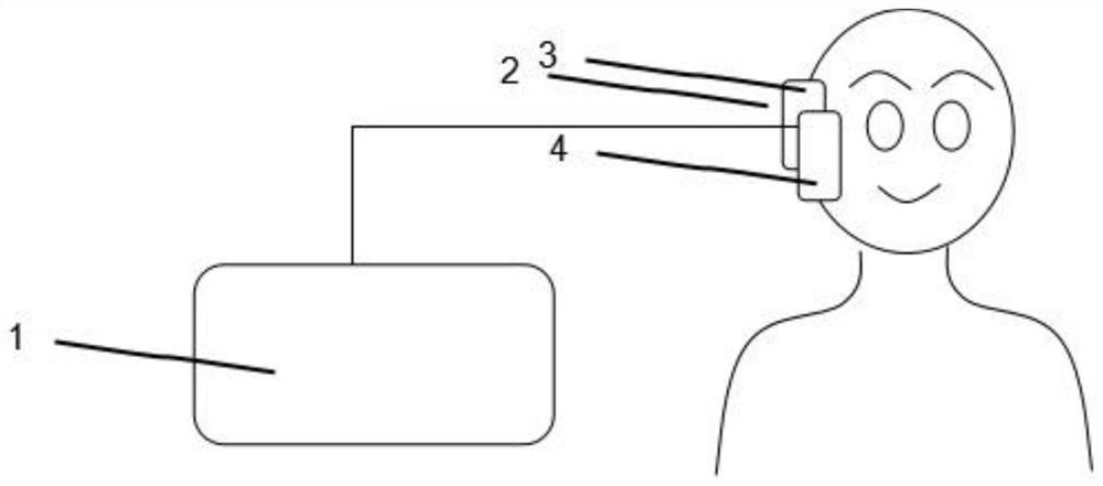
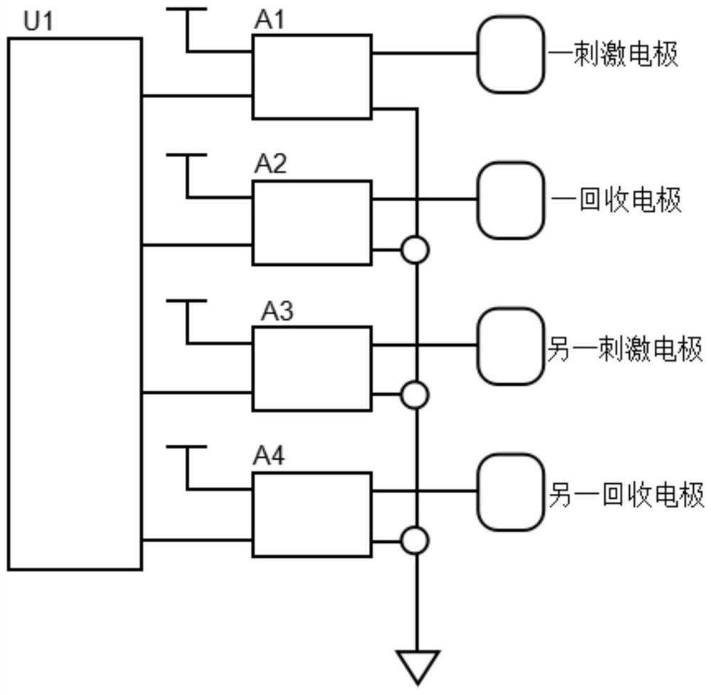
[0036] This embodiment: as figure 1 As shown, a retinal and optic nerve protective electrical stimulation device includes a stimulator 1, and the stimulation pulse output by the stimulator 1 is electrically stimulated on the nerve tissue on the visual conduction pathway through the amplifying electrode 2 across the retina or across the orbit.

[0037] The stimulator 1 outputs corresponding stimulation pulses by automatically controlling stimulation time and adjusting pulse waveform, pulse width, frequency, and amplitude.

[0038]Since the stimulation pulses output by the stimulator are electrically stimulated on the nerve tissue on the visual conduction pathway through the amplified electrodes, the preclinical research results using transcorneal electrical stimulation show that this treatment is also suitable for early post-injury patients. intervention, first proposed that transcorneal electrical stimulation was able to significantly increase the number of retinal ganglion ce...


PUM


Abstract

The invention belongs to the technical field of medical devices, and particularly relates to a retina and optic nerve protective electrical stimulation device, which is characterized in that stimulation pulses output by a stimulator electrically stimulate nervous tissues on a visual conduction path across the retina and eye sockets through an amplification electrode; the invention solves the problem of lack of research on non-invasive eye retina-crossing electrical stimulation, eye socket-crossing electrical stimulation and electrical stimulation devices thereof in the prior art, and has the beneficial technical effect of filling up the application blank of the non-invasive eye retina-crossing and eye socket-crossing electrical stimulation device.

Description

technical field [0001] The invention belongs to the technical field of medical devices, in particular to a retinal and optic nerve protective electrical stimulation device. Background technique [0002] Traumatic optic nerve injury can lead to retrograde apoptosis of retinal ganglion cells, and transcorneal electrical stimulation can increase cell survival. We monitored the morphology and survival of retinal ganglion cells after optic nerve injury in live animals by using retinal confocal methods. The optic nerve was injured in rats, and the number and shape of retinal ganglion cells were recorded before the injury and 3, 7, and 15 days after the injury. Three days after optic nerve injury, a large number of retinal ganglion cells survived in the stimulation group compared with the sham stimulation group by transcorneal electrical stimulation, the difference between the two groups became smaller at seven days, and disappeared at 15 days. Morphological analysis showed that ...

Claims


Application Information

Patent Timeline
IPC IPC(8): A61N1/36A61N1/04G05B19/042
CPCA61N1/0484A61N1/36014A61N1/36046G05B19/0423G05B2219/21085
Inventor 梁婷
Owner HARBIN MEDICAL UNIVERSITY
Learn More